Chapter 17
Clinicopathologic Correlates in Orbital Disease
ZEYNEL A. KARCIOGLU
Main Menu   Table Of Contents

Search

STRUCTURAL DISORDERS
ORBITAL INFLAMMATION
ORBITAL NEOPLASIA
OTHER ORBITAL DISORDERS
REFERENCES

Of all essential human senses, vision is the most important. The eye, organ of vision, should be well protected and supported functionally and structurally. This protection and support is provided by the orbit, which is a cone-shaped bony structure with a volume of 30 mL in which the 7-mL globe is positioned centrally and anteriorly. All the support systems of the globe, including the optic nerve, lacrimal gland, extraocular muscles, fibroadipose tissue, peripheral nerves, ganglionic tissue, and blood vessels are designed to be confined within approximately 25 mL of space surrounding the eyeball. Many tissues are crowded in this limited space and give origin to a variety of congenital, structural, inflammatory, degenerative, and neoplastic diseases. Further, orbital pathologies are not limited only to primary disorders; many systemic diseases and diseases of the globe may affect the orbit secondarily. The orbit is also secondarily affected by the diseases of the cranium, eyelids, conjunctiva, and the nasal cavity and paranasal sinuses. This chapter attempts to give an overview of the clinico-pathological correlation of orbital diseases. Because of limited space, entities that are encountered more often are detailed and some other less frequently seen pathologies are only mentioned with citations of pertinent references.
Back to Top
STRUCTURAL DISORDERS

CONGENITAL ANOMALIES

Orbitocraniofacial Deformities

The orbital bones begin to develop during the first 2 months of embryogenesis.1 Toward the end of the fifth week, the axes of the two orbits begin to move forward as a result of the growth of the maxillary processes.2 Most of the orbital bones are formed during the third month of gestation, but their complete ossification and fusing takes a longer time to complete, at approximately the seventh month of gestation. At term, the orbit is almost spherical in configuration. The periorbital sinuses begin to develop at approximately the second month of gestation and continue to grow after birth. Although the eye reaches adult size (approximately 7 mL) at approximately age of 2.5 years, the adult form and dimensions of the orbit (approximately 30 mL) are not finalized until puberty. Therefore, between the second month of embryogenesis and the age of puberty, numerous factors may affect the development of the orbit. For example, if the globe fails to develop or it is microphthalmic, the orbit does not attain its normal volume. However, if a congenital cyst or tumor develops within the orbit and expands its volume, the socket reaches a very large size unless the cyst is removed

Orbitocraniofacial deformities can be categorized in two large groups as craniosynostosis and clefting disorders.

Craniosynostosis

This developmental pathology of the orbit is dependent on the fusional abnormalities between the bones and it is divided into primary and secondary forms. Primary craniosynostosis refers to premature fusion of cranial sutures caused by embryologic error. Secondary synostosis is the premature closure of sutures because of other causes such as intrauterine trauma or effects of teratogenic drugs.3,4 Approximately 15% of craniosynostosis cases are associated with systemic abnormalities and are known as syndromic craniosynostosis; the remaining 85% represent nonsyndromic forms.5 Syndromic craniosynostosis represent a variety of inherited syndromes with abnormal development of the skull and orbit including: Apert syndrome with syndactyly of hands and feet, Crouzon syndrome with shallow orbits and proptosis, maxillary hypoplasia and cleft palates, and Pfeiffer syndrome with malformation of thumb and great toe and soft tissue syndactyly.6

Clefting Disorders

These malformations involve more extensive soft tissue abnormalities and depending on the location and the extent of the cleft, may be associated with bone malformations as well. The mapping system that is used to describe the location of a cleft is known as the Tessier clock face. In this scheme, the clefts are numbered from 0 to 14 beginning at the inferonasal area and moving clockwise to the superior nasal area. Commonly encountered clefting syndromes are: Goldenhar syndrome, Treacher-Collins syndrome, and the facial microsomias7,8 (Table 1).


TABLE 1. Summary of Orbito-Craniofacial Dysmorphic Syndromes


Syndrome Synonyms Manifestations*
Craniosynostosis syndromes
Crouzon syndrome Hereditary craniofacial dysostosis Premature sutural synostosis (coronal, sagittal, lambdoid sutures), shallow orbital cone with forward displacement of the globe, brachycephaly, hypertelorism, optic nerve atrophy, maxillary hypoplasia, parrot-beaked nose
Scheuthauer-Marie Sainton syndrome Cleidocranial dysostosis Brachycephaly, sutural synostoses, frontal bossing,maxillary hypoplasia, dental anomalies, aplasia of the clavicles and pubic bones
Acrosyndactyly syndromes
Apert syndrome Acrocephalosyndactyly I Premature cranial synostoses (beginning with the coronal sutures), brachycephaly, proptosis, hypertelorism, midface and maxillary hypoplasia, high palatal arch, prognathism, syndactyly of hands and feet
Carpenter syndrome Acrocephalopolysyndactyly Asymmetric craniosynostosis, polysyndactyly of the feet, agenesis of the middle phalanges, variable soft-tissue syndactylies, genu valgus, cardiac anomalies
Chotzen syndrome Saethre-Chotzen syndrome Craniosynostosis (variable, frequently asymmetric), low-set frontal hairline, ptosis, hypertelorism, mid-face asymmetry, brachydactyly, cutaneous syndactyly (2nd & 3rd fingers)
Pfeifer syndrome   Craniosynostosis with turribrachycephaly, slanted palpebral fissures, hypertelorism, hypoplasia of the midface and maxilla, dental anomalies, brachydactylies, soft-tissue syndactylies
Craniofacial deformities
Goldenhar syndrome Oculo-auricular vertebral dysplasia, Goldenhar-Gorlin syndrome, hemifacial microsomy Unilateral facial hypoplasia with hypoplasia of maxilla, temporal bone, and mandible, epibulbar epidermoid and coloboma of the upper lid, microphthalmos, anomalies of the ear, vertebral anomalies
Treacher-Collins syndrome Franceschetti syndrome Bilateral hypoplasia of the zygomatic bone, temporal bone, maxilla and mandible with typical “bird face,” antimongoloid slant of the palpebral fissure, coloboma of the lid, abnormalities of the ear, cleft palate
Robin syndrome Pierre Robin sequence Hypoplasia of the mandible, cleft palate, glossoptosia

*Ocular & adnexal manifestations are listed in bold print.


ANOPHTHALMOS/MICROPHTHALMOS

When the globe is abnormally developed, microphthalmos, congenital cystic eye, and extremely rarely, anophthalmos occur. Microphthalmos usually occurs as a unilateral condition and in approximately 10% of cases it is associated with other craniofacial malformations including agenesis of the corpus callosum, polymicrogyria, and mid-line arachnoidal cysts. Microphthalmos may be seen as a part of several genetically determined neuronal migration disorders such as Walker-Warburg syndrome, Aicardi syndrome, and Fukuyama congential muscular dystrophy.9,10

In cases of microphthalmos and anophthalmos the orbit may be well formed but does not develop to a full adult volume. The mechanism by which the presence of the globe effects the growth of the orbit is not well understood. Microphthalmos may be associated with a colobomatous cyst as a result of the abnormal closure of the embryonic optic fissure leading to the prolapse of neuroectodermal tissues into the orbit (Fig. 1) This cystic structure may increase rapidly in size to overshadow the abnormal globe and may be confused with a neoplasm. When cystic lesions in the orbit are suspected imaging studies should be performed not only to look for other intracranial abnormalities but also to establish the possible connection of the cyst to the colobomatous globe versus to abnormally formed meninges.11 Macrophthalmos (buphthalmos) may also rarely develop as a congenital anomaly in patients with Sturge-Weber syndrome and rarely in neurofibromatosis type I.

Fig. 1 Congenital lesions. A very large cystic teratoma of a 1-month old child (A). B. Histology of this lesion that contains a variety of tissues, endodermal, ectodermal, and mesenchymal. An orbital cyst (C) in an orbit containing micro-ophthalmic globe. C, D. The protrusion of the cyst inferiorly creates a mechanical lower lid ptosis that narrows the right maldeveloped conjunctival sac even further. Axial and sagittal CT scan showing a meningoencephalocele (ME) occupying the entire orbit (E). The histopathologic examination of the lesion revealed both meningeal (M) and brain (B) tissues (F). The high-power histopathology reveals ciliated ependymal cells lining some of the cystic spaces (arrowhead) (G).

ORBITO-CRANIAL MALDEVELOPMENTS

Cephalocele results from the extension of maldeveloped CNS tissues including meninges (meningocele), brain parenchyma (encephalocele), and the combination of the two (meningoencephalocele) into the orbital cavity.12,13 Intraorbital cephaloceles may develop anteriorly at the suture lines of orbital bones or posteriorly extending into the orbit from orbital fissures and the optic canal. Depending on the combination of these herniations they contain brain and/or meningeal tissues (Fig. 1). Aberrant fibroglial tissue has also been described in the orbit.14

Hamartoma is a tumor-like proliferation of tissues that normally exist at a given body location. The best examples of orbital hamartomas are the vascular hamartomatous lesions that are composed of vascular elements including capillary endothelial cells, distended or collapsed cavernous blood and lymph vessels, tortuous arterial and venous channels with or without anastomoses, etc. Other examples of hamartomatous orbital tumors include neurofibroma and lipomatous hamartoma.15

Choristoma, however, is a tumor-like proliferation of tissues that are not normally present at a given body location. The most commonly encountered example of orbital choristoma is a dermoid.16 Dermoids that present with many varieties result from the entrapment of epithelial structures at the site of closure of fetal fissures. Superficial dermoid cysts occur primarily subcutaneously anterior to orbital septum or within the anterior orbit. If the cyst wall is made of epidermis without dermal tissues, it is classified as an epidermoid cyst. These lesions are occasionally lined by conjunctival or pseudostratified respiratory epithelium.17 The superficial lesions must be distinguished from deep orbital dermoids that are usually rounded, encapsulated tumors filled with fatty materials, keratin, and dermal structures such as hair particles. Histopathologically the dermoid wall is lined by keratizing squamous epithelium with dermal appendices including hair follicles and sebaceous and eccrine glands.18

Most of the dermoids are well outlined by ultrasonography because of their anterior location thus making CT or MRI rarely necessary.19 If the dermoid is unusually large or located at the frontal zygomatic suture, CT is necessary to document the relationship of the lesion to the bone before surgical intervention. Rarely, dermoids at the fronto-zygomatic suture may develop dumbbell-shaped lesions partially within the orbit and partially extending into the temporal fossa.20,21 Unusually large superior orbital dermoids particularly those that leak and create granulomatous reaction within adjacent soft tissues may erode the bone and extend into the frontal sinus or the cranium (Fig. 2).

Fig. 2 Dermoid. Different presentations of dermoid (d): superior medial, semi-solid mass pushing the globe down and out (A); a ruptured dermoid causing an inflammatory reaction within adjacent soft tissues (B); a large superior lateral dermoid eroding through the roof of the orbit to extend into the brain (C); extraorbital dermoid within the subcutaneous tissues of the eyebrow (D); gross appearance of the cystic dermoid containing whitish yellow cheesy keratin material intermixed with hair (E); dumbbell dermoid that is present on both sides of the frontozygomatic fissure (F); histopathology of dermoid wall (dw) containing skin appendages, the lumen of the dermoid is lined with stratified squamous epithelium producing keratin (K) (G). ([E] is the courtesy of Amin M. Nasr, MD of Beirut, Lebanon)

ORBITAL TERATOMA

Teratoma is a germ-cell tumor that contains tissues derived from endoderm, ectoderm, and mesoderm22 (Fig. 1). Therefore, these lesions may contain skin, bowel, lung, brain, thyroid, cartilage, and bone tissues. Most teratomas develop unilaterally and in girls. A majority of these congenital tumors are benign. Occasional reports have documented malignant transformation within orbitocranial teratomas.23 However, these benign tumors continue to grow after birth because of the collection of secretions from different tissues into the partially cystic spaces of the tumor. Some teratomas create massive proptosis and most can only be treated by exenteration. However, some of these lesions have recently been reported to be removed surgically with preservation of the globe and other vital orbital structures.

TRAUMA

Mechanical Injury

Orbital injuries result from the absorption of kinetic energy that occurs whenever the orbital tissues contact an object moving at a different speed.25 The orbital rim is capable of absorbing a considerable amount of kinetic energy without being fractured. Yet, a variety of impact forces striking the orbit may result in fractures in different areas.26 The absorption of the kinetic energy by an orbital bone may lead to contusion and/or laceration of the skin and superficial soft tissues, local deformation of the adjacent structures, globe, orbital soft tissues and bones and increases pressure in the orbital cavity. A common end result of an orbital impact is the fracture of the floor and/or the medial wall (lamina papyracea)27 (Fig. 3). Fractures of other orbital bones occur less often. Foreign bodies may be introduced into the orbit at the time of injury and may cause secondary problems depending on the nature and the location of the foreign body.28 Some foreign bodies such as copper may cause tissue necrosis and degeneration (chalcosis), and others particularly organic matter, may carry organisms such as bacteria and fungi into the orbital tissues and cause secondary infections29 (Fig. 3). Once the fracture of an orbital bone occurs, it may produce sharp edges to lacerate adjacent soft tissue structures including the globe, optic nerve, other nerves, muscles, and vessels.30 Depending on the damage of the particular tissue, functional deficit results.

Fig. 3 Orbital trauma. Axial CT scan (A) and T1-weighted MRI (B) show multiple organic foreign bodies (fb) within the left medial rectus muscle and posterior orbit. The round tissue depicted in the inset was removed at the time of surgery; histopathologically it proved to be a foreign body granuloma (fbg). A coronal CT scan (C) and the intraoperative photograph (D) depict a large, inferior orbital rim (ior) fracture. The right inferior rectus muscle that was prolapsed into the maxillary sinus is highlighted with an arrowhead (C). (A and B are the courtesy of J. Christopher Fleming, MD of Memphis, TN)

Another issue to deal with in an injured orbit is the development of hematoma, hematic cyst, and cholesteotoma. Hemorrhage in the orbit may occur spontaneously without any physical exertion in healthy individuals. Although terminology is not very strict, hematoma usually refers to a localized collection of blood within orbital soft tissues that develops secondary to trauma. When the blood collection within the orbit becomes organized and surrounded by a thin pseudocapsule, it is known as a hematic cyst31 (Fig. 4). If the hemorrhage develops within an existing lymphatic or vascular tumor, these lesions are known as blood cysts or “chocolate” cysts.32

Fig. 4 Hematic cyst. Axial CT scan (A) showing a large superiorly located, well-circumscribed hematic cyst (hc) presenting as an homogeneous low-density image. Intraoperative photograph of the same case shows dark brown hematic cyst. (B). C and D show the gross and histopathologic appearance of the hematic cyst respectively. It is surrounded by a fibrous pseudocapsule (arrows) containing a mixture of cholesterol crystals, (cc), hematoidin crystals (HC), and other proteineceous debris.

Hematic cyst consists of a localized collection of blood surrounded by a nonepithelium-lined thin fibrous capsule.33 These lesions usually develop within 1 to 2 weeks of orbital trauma but chronic cases may occur up to 20 years after orbital injury.34,35 They may reach to a size causing proptosis, extraocular motility disturbance, compression on the globe and optic nerve, that can easily be detected with ultrasonography, CT, or MRI. Hematic cysts may develop within the muscle cone or in the extraconal orbital locations.33–36 These cysts are lined by fibrovascular tissue at the periphery and contain degenerated erythrocytes, protein debris, and cholesterol crystals. In many instances the thin nonepithelial lining is adherent to the adjacent structures with fibrous tissue.

Cholesteatoma is another cystic lesion that is confined within a “pseudowall” without an epithelial lining.37 Cholesteotomas are usually located in the superior lateral orbit within the lacrimal gland fossa. Imaging studies may show a cystic, semi-cystic, or a solid lesion within the diploe of the bone or within the orbital soft tissues, with or without erosion of the adjacent bone.38 Histopathologically the lesion is composed of cholesterol clefts, hemosiderin, and hematoidin granules, other blood breakdown products and fibrin surrounded by a mixed lymphohistiocytic infiltrate and multinucleated foreign body giant cells.39

On imaging studies these lesions appear as unilocular rounded masses with destruction of the adjacent frontal and zygomatic bones. Although bone involvement in general implies malignancy, the sclerosing character of the bony destruction in choleosteoma, which is best seen in bone window images, favors a benign lesion. Although bone destruction also makes one think along the lines of metastatic tumors, one should also consider benign lesions such as brown tumor, aneurysmal bone cyst, and ruptured dermoid. Multiple cuts of the frontal bone should be examined to rule out the possibility of intracranial extension.

Osteomyelitis of the orbital bones evolving as a complication of paranasal sinusitis is another entity that should be considered in the differential diagnosis of cholesteotoma. In osteomyelitis the bone infection extends into the periosteal space and beyond. Precise delineation of the lesion can be performed with CT and MRI particularly in combination with bone SPECT, a sensitive technique used to detect osteomyelitis within cranial and orbital bones.40

Mucocele

Although a commonly encountered space occupying lesion in the orbit, mucocele is technically not a neoplasm. It is a cystic cavity lined by pseudostratified respiratory epithelium prolapsing into the orbit from a paranasal sinus, most commonly the frontal followed by the ethmoidal sinus (Fig. 5). Primary mucoceles develop as a result of an inflammatory obstruction of the ostium of the paranasal sinuses. Secondary mucoceles, however, are most commonly seen after orbital trauma and surgery; they may also develop secondary to neoplasms of paranasal sinuses and nasopharynx. If there is a superimposed infection, the lesion is referred to as pyocele. The mucocele develops as a well delineated cystic structure originating from a paranasal sinus. Depending on the location, it may compress orbital structures including extraocular muscles, optic nerve, and the globe.41 Clinical presentation of the mucocele is usually with globe displacement and/or proptosis, extraocular motility deficiency, particularly in the direction of the sinus extension into the orbit, and other compressive symptoms.42 The crepitant or calcified hard wall of the mucocele may be palpated underneath the superior or medial orbital rim. Mucoceles in general, are rare in children, however, a unique variant, ethmoidal mucopyocele, is known to occur in the medial canthal area, with lateral displacement of the globe.

Fig. 5 Mucocele. Moderate proptosis and slight lateral displacement of the left eye secondary to a large medially located mucocele (m) originating from the ethmoid sinus (A,B). Note the compression of the calcified wall of the lesion onto the globe and the optic nerve (B). The large cystic nature of the mucocele with low internal reflectivity and segmentally calcified wall is demonstrated by ultrasonography (C). Gross specimen of the stripped mucosal lining of the mucocele from the same case (D).

On CT, mucoceles present as hypointense, expanding masses originating from the paranasal sinuses. Early in their development these lesions are small, mucous-containing cysts. Later they are characterized by crescent-shaped and thinned remodeling of the bony walls of the orbit and sinuses.43 On MRI, mucocele presents with different appearances depending on the amount of free water within its luminal contents. When the intraluminal mucous becomes inspissated, the signal intensity in both T1 and T2 images decrease, getting closer to normal air content of the sinus.44 Treatment of mucocele is surgical excision.

Other injuries with toxic chemicals and radiation are known to damage orbital tissues.45–47

VASCULAR MALFORMATIONS

Arteriovenous Fistula

Orbital arteriovenous (AV) fistulas are established as a result of abnormal flow between the arteries and veins. These lesions can be divided into three basic types: carotid cavernous, dural and orbital AV fistulas. Carotid cavernous fistula is usually traumatic but may also develop secondary to a rupture of an aneurysm particularly in elderly atherosclerotic patients. These fistulas commonly develop between an intracavernous segment of internal carotid artery and cavernous sinus and shunt arterial blood into superior ophthalmic vein.48 Dural cavernous fistulas, however, develop between small meningeal branches of internal/external carotid artery and the cavernous sinus. These small vessels that have thin walls that may rupture spontaneously particularly in hypertensive individuals, secondary to minor trauma and maintain a low blood flow.

Orbital AV fistulas usually develop secondary to traumatic rupture of the ethmoidal artery into the orbital venous system. This type of fistula maintains a low blood flow. Clinical findings of AV fistulas include rapidly developing proptosis, edema of the conjunctiva and eyelids, dilatation and tortuosity of the conjunctival and episcleral vessels, and secondary glaucoma.

Most of these cases are diagnosed with imaging procedures including CT, MRI, angiography, color Doppler ultrasonography, and catheterized angiography.49 Current treatment of these lesions is embolization via catherization.50 Morphologic data are limited to autopsy material because most patients with AV fistulas do not undergo biopsy procedure. These lesions show irregular, malformed arteries and veins with abnormal elastic and muscular layers and secondary endothelial cell proliferation. Approximately half of the low shunt fistulas close spontaneously;51 therefore, it is best to follow-up some of these patients conservatively if they do not have severe symptoms.

Orbital Varix

Orbital varix is a rare vascular lesion with questionable histopathogenesis. The absence of valves in the orbital venous system and the weakening of venous wall may lead to pooling and stasis of blood resulting in distention of the venous channel with thrombosis. In gross appearance, the varix is a distended vein containing a canalized or uncanalized thrombus.52,53 Histopathologically varix consist of irregular vascular channels lined by endothelial cells. In chronic lesions, the blood vessel walls irregularly thicken with fibrosis and deposits of chronic inflammatory cells mixed with deposits of calcium and hemosiderin pigment are seen. Orbital varices are divided into primary and secondary types. The primary orbital varix is confined to the orbit as an isolated lesion without any connection to other A-V malformations. The secondary orbital varix, however, develops as an extension of an intracranial AV malformation that shunts blood to the orbital venous system causing the venous channels to distend secondarily.54 Management of orbital varix consists of total surgical excision when possible and/or endovascular embolization.

Back to Top
ORBITAL INFLAMMATION

MICROBIAL INFLAMMATION

Microbial infections of the orbit develop because of a variety of organisms, which are introduced to the orbit by different routes. Bacterial orbital infections are the most common that primarily originate from the paranasal sinuses and to a lesser degree, secondary to penetrating injuries with or without foreign bodies and rarely, secondary to endophthalmitis or systemic infectious process such as bacterial septicemia, tuberculosis, etc. The most common cause of bacterial orbital cellulitis is Staphylocci and Streptococci species. Although Haemophilus influenza was a common causative organism in children younger than 4 years of age, recent vaccination efforts have decreased the incidence of H. influenza cellulitis significantly.55

Anatomically speaking, if the infection is limited to anterior of the orbital septum, involving the eyelids it manifests itself primarily with lid and conjunctival edema and some venous congestion but not with proptosis.56 This process that is known as preseptal cellulitis is usually initiated with skin injuries or an upper respiratory tract infections in children. However, if the infectious process involves orbital soft tissues behind the septum, it is called orbital cellulitis (Fig. 6). Cellulitis usually develops secondary to a paranasal sinus disease, clinically associated with axial proptosis, marked chemosis, congestion of retinal veins, limitation of extraocular motility, and swelling and pain around the eye. Depending on the severity of the condition, vital structures within the orbit such as the globe and the optic nerve may be compressed or infiltrated by the infection. Generally, in children the orbital cellulitis originates from the ethmoid sinus and may limit itself to the subperiosteal site for a while and later spread into orbital soft tissues.

Fig. 6 Orbital cellulitis. A and B show the appearance of right orbital cellulitis secondary to Staphylococcus infection from right ethmoidal sinus. The proptosis, chemosis and visual loss in this patient worsened after the ethmoidectomy and the medial orbit had to be explored with additional drainage of pus. Note the stretch of the right optic nerve and pear-shaped right globe secondary to marked proptosis on an axial CT scan (B). C shows another case with severe ethmoiditis causing cellulitis of the right orbit with the extension of the infection preseptally toward the left globe. The patient responded well to ethmoidectomy and drainage of the abscess from the right medial orbit. D shows the appearance of a preseptal and anterior orbital cellulitis secondary to a long-standing foreign body caused in a motor vehicle accident. The patient depicted in frame E developed Streptococcal orbital cellulitis with secondary brain abscess as depicted in the sagittal T1-weighted MRI (F).

Acute bacterial orbital cellulitis presents a nonspecific histopathology associated with diffuse polymorphonuclear infiltrate that may on occasion lead to abscess formation57 (Table 2). Subperiosteal and soft tissue abscesses may present with decreased vision, disc edema and increased intraorbital and intraocular pressures. Orbital abscess is best demonstrated with MRI. Emergent orbital exploration and the drainage of the abscess is indicated.


TABLE 2. Correlation of Cellular Pathology to Clinic Diagnosis in Orbital Inflammatory Lesions


AcuteSubacuteChronicLympho-proliferativeGranulomatousVasculitis
PMNs, necrosis, eosinophils, cellular debris, w/ or w/o organismsPMNs, lymphocytes, plasma cells, histiocytesLymphocytes, plasma cells, histocytes, fibroblastsLymphocytes. w or w/o germinal centersLymphocytes, giant cells, histocytes w/ or w/o focal necrosis w/ or w/o organismsAcute and/or chronic inflammatory cells around blood vessels, blood vessel necrosis w/ or w/o giant cells
Cellulitis, acute IOI, Sjogren, sino-orbital aspergillosisResolving cellulitis, subacute IOIChronic IOI, underlying neoplasmIOI, lymphocytic or atypical lymphocytic hyperplasia, low grade B-cell lymphoma, lymphangioma, non-specific inflammation, SjogrenForeign body, sarcoidosis, tuberculosis, fungal infections, IOI, ruptured dermoid, fat necrosis (lipogranuloma)giant cell arteritis, Wegener, polyarteritis, mucormycosis

IOI, idiopathic orbital inflammation (orbital pseudotumor); PMN, polymorphonuclear cell; w, with; w/o, without.


Orbital cellulitis may also be caused by different types of foreign bodies including organic and nonorganic matter and nonautogenous surgical implants.28 Inorganic foreign materials, such as metal and glass, are usually well-tolerated and do not cause infection unless they significantly distort the orbital anatomy with exposure to periorbital sinuses and nasal cavity. Organic foreign bodies such as wood, vegetable fibers, etc., however, trigger significant foreign body reaction and sometimes suppurative inflammation.These should be documented by CT and/or MRI and surgically removed29 (Fig. 3). Today many nonautogenous materials are used in ocular and orbital reconstruction, including porous implants, mesh materials, polymeric silicone plates, sponges for scleral buckling procedures and reservoirs of drainage valves for glaucoma. Implants and repair blocks made of porous materials may lead to acute infection when they erode through sinus or conjunctival epithelium. Whenever there is a foreign body, noncaseating granulomatous reaction with multinucleated giant cells is identified, adjacent to the foreign material; secondary acute inflammation may be superimposed. In penetrating injuries the nature of the foreign body is an important factor.

An exception to suppurative soft tissue reaction that is caused by bacteria is chronic caseating granulomatous inflammation, which is caused by mycobacteria and certain types of fungi (Table 2). Ocular and adnexal tuberculosis is usually seen with manifestation of systemic mycobacterial infection,58,59 Because of the recent increase in the numbers of immunologically suppressed individuals secondary to viral epidemics and wider use of immunosuppressant antimetabolites in longer-surviving cancer and transplantation patients, the incidence of tuberculosis has been increasing steadily during the past two decades and the clinical picture of the disease has been changing, with many cases developing because of atypical mycobacteria that are resistant to traditional multidrug treatment.61 It has been reported that the individuals with HIV/AIDS have an incidence of tuberculosis 500-fold more than that is seen in the general population.62

The orbital disease, is more often seen in children and nonwhite patients. History of antecedent penetrating injury is a common presentation of tuberculosis, caused by atypical mycobacteria. Histopathology of tuberculosis consists of zonal granulomatous inflammation with numerous epitheliod histiocytes surrounding a necrotic (caseating) center. Tissue diagnosis is pathognomonic only with the documentation of positive acid-fast organisms; however, in many cases special stains may fail to demonstrate the mycobacteria, but the cultures grow M. tuberculosis or atypical mycobacteria. Orbital tuberculosis is usually associated with systemic disease (Fig. 7).

Fig. 7 Orbital tuberculosis. A 2-year-old boy with left orbital tuberculosis (A). Both mother and child had systemic disease. A case of bilateral orbital and perinasal sinus tuberculosis is shown in the coronal CT of a 28-year-old man (B). Note the irregular involvement of the bony tissues of the orbits and the sinuses. C and D show a caseating granuloma (c) and AFB-positive tuberculous bacilli in necrotic inflammation (arrowheads) respectively.

In most instances, fungi infect the orbit as an extension of paranasal sinus disease or after penetrating injury associated with the introduction of organic matter. Most of the fungal infections, particularly mucormycosis often develop in immunocompromised patients.63

Orbital mucormycosis is an emergency situation because it causes rapidly progressing necrotizing inflammation secondary to vascular involvement (Fig. 8). Orbital exploration should be performed immediately to establish the diagnosis by identifying the broad, nonseptated hyphae and for surgical debridement as well as irrigation with antifungal agents. The prognosis of mucormycosis is very poor. Aspergillosis, unlike mucormycosis, presents a low-grade, smoldering chronic granulomatous inflammation that may be confused with primary orbital tumor.64 The identification of the organism in fungal and parasitic disease, is crucial at the time of surgery; fungi may also be identified with smears and frozen sections.65 In any kind of orbital exploration secondary to cellulitis, tissue samples should be obtained for Gram, fungal and AFB stains and aerobic, anaerobic and fungal cultures.

Fig. 8 Mucormycosis. The appareance of mucormycosis in the right orbit, periorbital skin and maxillary sinuses of a 60-year-old man with diabetic ketoacidosis (A). B shows the funduscopic apperance of a central retinal artery thromboembolism with resultant “cherry-red spot” from another case of orbital mucormycosis. C shows the extensively necrotic, bloodless cut surface of the exenteration specimen from the patient shown in A. Histopathologic examination of this specimen revealed numerous nonseptated mucormycosis hyphae with 90-degree branching (D) in Gomori methanamine silver (GMS) stain.

As a rule, no matter how rarely, any microbial inflammation, including bacteria, fungi, viruses, and others, may infect the orbit and cause acute, chronic or granulomatous inflammation leading to tissue damage and fibrosis. These rare diseases include syphilis,66 leprosy,67,68 Parinaud syndrome,71,72 and actinomycosis.73,74 Examples of parasitic orbital infections include echinococcosis, cysticercosis, myiasis, and trichinosis of extraocular muscles.76–81

NONMICROBIAL INFLAMMATION

Many forms of inflammatory processes may trigger orbital inflammation that simulates neoplasms by producing proptosis and associated orbital findings.82 These include Graves disease,83–85 idiopathic orbital inflammation (orbital pseudotumor),86,87 Tolosa-Hunt syndrome,88,89 sarcoidosis,90,91 Sjögren syndrome,92,93 and Wegener granulomatosis.94–96

Graves Disease

Thyroid-associated orbitopathy, better known as Graves disease (Gd), is an idiopathic orbital inflammation that primarily involves the muscles and soft tissues of the orbit and the eyelids. The commonly involved muscles include inferior, medial, superior and lateral recti that cause swelling of the tissue leading to proptosis and eyelid retraction (Fig. 9). Gd is the most common cause of unilateral and bilateral proptosis in adults; although uncommonly it may be seen in children as well.

Fig. 9 Graves disease. Clinical findings of Graves disease are depicted in frame A, including bilateral proptosis and lid lag with extraocular motility disturbance, chemosis with prolapse of lacrimal gland and congestion of conjunctival and episcleral blood vessels. Axial CT scan (B) reveals marked swelling of all recti muscles. The histopathologic appearance of the extraocular muscle from a patient with Graves disease reveals chronic inflammatory infiltrates, primarily composed of lymphocytes and plasma cells (C). The extraocular muscle volume is increased because of diffuse endomysial fibrosis, mucopolysacccharide deposition and chronic inflammatory cell infiltration (D). The orbital fat, meninges and the optic nerve (blue arrow), large blood vessels of the orbit, such as ophthalmic artery and its branches (green arrows) and the ciliary ganglion (red arrow) do not show any inflammation. The enlargement of the extraocular muscles are well depicted in frame E, which represents a transverse section, approximately at the level indicated by the yellow line in frame B. (LR: lateral rectus; SR + L: superior rectus and levator complex; SO: superior oblique; MR: medial rectus; IR: inferior rectus; IO: inferior oblique). (Frames C, D, and E are the courtesy of Ralph C. Eagle, MD of Philadelphia, PA)

The pathogenesis of Gd is not completely understood; therefore, it is labeled as an “autoimmune” process.82,98 It has been suggested that individuals with HLA-B8 major histocompatility antigen Haplotype are genetically susceptible to Gd.99 The hypothesis is that circulating T-cells directed against an antigen in thyroid follicular cells recognize a similar antigen in extraocular muscles and orbital soft tissues.100 Experimental studies suggest that thyrotropin receptor (TSH-R) is one of the possible entities to stimulate the autoantibodies that lead to the inflammatory changes within orbital soft tissues. In Gd there is a predominance of T-cells with Th1 profile, although Th2 profile of cytokine production has also been reported.100 The cytokines stimulate fibroblasts to produce glycosaminoglycans that in turn lead to deposition of this substance within the muscle tissue leading to anatomic and functional deficiencies (Fig. 9). Although cell mediated immune reaction predominate in early Gd, humoral immunity plays a greater role in later phases.101

Some of the immune changes are reflected in the histopathology of Gd, which can basically be divided in two stages. The active inflammatory stage consists of perivascular edema and clustering of lymphocytes and plasma cells; lymphoid follicles are not frequent in Gd. In the chronic stage the volume of the involved orbital tissues are increased because of the deposition of glycoproteins and mucopolysaccharides and secondary to the infiltration of fibroblasts producing collagen. Later in chronic stages of the disease, the edema decreases and the muscles are primarily infiltrated with interstitial fibroblasts and chronic inflammatory cells leading to fibrosis.

Although 80% of patients with Gd present with a history of hyperthyroidism, approximately 10% suffer hypothyroidism or autoimmune thyroiditis; occasionally, an euthyhroid individual may also develop the signs and symptoms of Gd.102 Upper lid retraction is the most frequent clinical sign in early Gd (75%) followed by asymmetrical bilateral proptosis (60%) and restriction of extraocular muscles (40%). Compressive optic neuropathy may result secondary to the enlargement of the extraocular muscles in the apex. Optic nerve malfunction is manifested by afferent pupillary defects and color vision and visual field deficiencies in approximately 5% of Gd patients.

The best means of evaluation of extraocular muscles is imaging with CT and/or MRI.11 Axial CT scan is very valuable to depict the enlargement of the extraocular muscles and determine whether there is any infiltration into other orbital soft tissues. Coronal sections are also very useful to evaluate the enlargement of the muscles and their relationship to the optic nerve in the orbital apex.103 In differential diagnosis of Gd, one should keep in mind that it is not only the most frequent cause of bilateral proptosis but unilateral proptosis as well. Therefore, the slowly progressive unilateral presentation may be confused with orbital pseudotumors, neoplasia, and solitary vascular lesions such as orbital varix.104,105 Orbital metastatic neoplasms may also be confusing if they are limited to the extraocular muscles. Imaging usually reveals nodular enlargement of the muscle and the diagnosis of a metastatic disease may be confirmed with fine needle aspiration biopsy (FNAB). The treatment of Gd includes oral and intravenous steroids, radiation and surgery.

Idiopathic Orbital Inflammation (IOI)

Orbital pseudotumor is a nonspecific chronic inflammatory condition of unknown etiology. Although an underlying immune process is suspected, no conclusive mechanism has been established for the development of this curious entity.87 IOI may develop with sudden onset of painful proptosis associated with motility disturbances, eyelid swelling, redness and chemosis. It may develop as a diffuse or localized lesion and its histopathology varies accordingly from case to case; the histopathology is also variable at different stages of the disease. The extraocular muscles may be involved with the inflammatory process but the main target is the orbital fibroadipose tissue. The inflammatory process may be grouped into two main categories: (1) diffuse and (2) localized nonspecific orbital inflammation. The localized nonspecific inflammation is further divided according to specific sites, i.e., myositis, dacryoadenitis, periscleritis and perineuritis. Each of these subgroups may present as an acute, subacute or chronic process in a given patient.

The histopathology of the IOI usually consists of a mixed polymorphonuclear and lymphocytic infiltrate during the early phases; as the disease advances, lymphoid follicle formation and fibrous tissue proliferation dominates the picture106 (Fig. 10). Patchy aggregates of lymphocytes and/or lymphoid follicles are frequently seen, but these lymphoid aggregates are not confluent as in lymphoid neoplasia and hyperplasia. Because of the diffuse fibrous reaction and the nonspecific nature of the mixed inflammatory infiltrate, the biopsy diagnosis of pseudotumor is not pathognomonic, but it should be correlated with clinical and radiologic findings in each case. Histopathologic patterns may vary in different regions of the specimen, therefore, it is advisable that these biopsies are processed totally. Hard granulomas, perivascular lymphocytic infiltrates, and occasionally true vasculitis may be identified within the orbital tissues of clinically typical IOI. Although polymorphonuclear leukocytes and eosinophils are occasionally seen, prominent acute inflammatory infiltrates should lead the pathologist to consider a vasculitis, such as polyarthritis nodosa or Wegener granulomatosis.

Fig. 10 Orbital pseudotumor. A patient with subacute orbital pseudotumor showing mild proptosis with hyperemia and edema of the eyelids and limited extraocular motility of the left eye (A, B). The left medial rectus muscle is severely involved with the disease that is also present to a lesser degree within the left inferior rectus muscle. Frames C and D reveal a mixture of lymphocytes, plasma cells, and eosinophils wthin fibroadipose tissues of the orbit.

Tolusa-Hunt syndrome, otherwise known as painful external ophthalmoplegia, is another inflammatory process of the orbit of unknown etiology. It is conceivable to think that it represents a localized form of idiopathic orbital inflammation (IOI) and presents with typical clinical manifestations because of its presentation in the orbital apex. The clinical symptoms include a severe, constant deep orbital pain associated with functional deficiencies of third, fourth, fifth and sixth cranial nerves.107 Typically, the orbital pain that presents abruptly also responds to systemic corticosteroid treatment with the same abruptness. Other symptoms of the disease including third, fourth, and sixth cranial nerves palsies and the hypesthesia of the periorbital skin also respond well to corticosteroid treatment. Although bilateral cases do occur; a great majority of patients with Tolosa-Hunt syndrome present unilaterally and therefore, should be differentiated from the tumors, which may involve the orbital apex including meningioma, pituitary adenoma, neurofibroma, paraganglioma, nasopharyngeal squamous cell carcinoma and metastatic tumors.108,109

Tumors of the apex, however, usually cause a gradual development of motility dysfunction depending on the location of the tumor that may be accompanied with dull pain but usually not with an abrupt onset of panophthalmoloplegia and explosive pain.

Nonspecific Granulomatous Inflammation

Sarcoidosis is a multisystem disease of an idiopathic nature that commonly involves the orbit and the eye. Systemically it involves the lungs and the upper respiratory tract, liver, spleen, lymphatic and hematopoetic tissues, central nervous system and the skin. Although there is considerable evidence in favor of sarcoidosis being infectious in nature, no causative agent has been established so far. The etiopathogenesis of sarcoidosis is still unknown.110 The typical noncaseating granulomas are made of T-lymphocytes of helper and suppressor types and dendritic Langerhans cells with human leukocyte antigen (HLA)-DR expression. Perivascular inflammatory reaction is characteristic of a delayed type hypersensitivity response. Some investigators think that noncaseating epithelioid granulomatous reaction, which is determined as the hallmark feature of the disease may be the host's immune response to presently unknown causative agent(s). Although the exact significance of granuloma formation in sarcoidosis is not clearly known, it appears that this tissue reaction is a secondary event as a result of exaggerated cellular immune response to a class of unknown antigens. It is hypothesized that the suppressor aspect of cell mediated response in sarcoidosis is abnormal and therefore reduces the function of helper T-lymphocytes. The initial step in granuloma formation of sarcoidosis is considered to be triggered by a cytokine (interleukin-1) that increases the proliferation of helper T-lymphocytes and activate these cells. Activated helper T-cells in turn secrete interleukin-2; a mitogen, which stimulates the proliferation of helper T-cells even further.111 As a consequence these cells aggregrate at the site of the causative insult and secrete monocyte chemotactic factors that lead to the gathering of epitheloid macrophages and multinucleated giant cells to form granulomas. Sarcoidosis is also associated with increased B-cell activity manifested by polyclonal hyperglobulinemia.112–114

Sarcoid granulomas are made of epithelioid cells and multinucleated giant cells, surrounded by lymphocytes and occasional plasma cells (Fig. 11). Many inclusion bodies have been described in the giant cells of sarcoidosis but none of these is pathognomonic. The granulomatous response of sarcoidosis is rather typical but not unique for this entity; fungal diseases, tuberculosis, Crohn disease and leprosy may produce similar granulomas115 (Table 2).

Fig. 11 Sarcoidosis. The appearance of a large sarcoidosis granuloma involving the anterior orbit and periorbital skin (g). The central dome-like lesion is caused by secondary to a Staphylococcus infection. In addition, plaquoid skin lesions of sarcoidosis are seen above the left eyebrow, on the left upper eyelid, and on the periorbital skin (A). The T2-weighted axial MRI with contrast reveals bilateral enlargement of lacrimal glands (g) in another case B. Frames C and D show T1-weighted sagittal MRIs with localized granulomatous masses of sarcoidosis within the posterior orbit involving the apex and extending into the cavernous sinus (C, D). A well-delineated but not encapsulated mass of a sarcoid granuloma (E). The histopathology of sarcoidosis consists of multiple granulomas composed of histiocytic cells, chronic inflammatory cells, and multinucleated giant cells (g) (F).

Approximately one-fourth of sarcoidosis patients develop ocular and orbital manifestations including anterior and posterior uveitis, chorioretinitis, conjunctival and eyelid granulomas and orbital mass lesions (Fig. 11). Lacrimal gland is a common site of involvement; autopsy studies show a high percentage of microscopic disease; however, only 15% to 20% of the patients show clinical symptoms. Although virtually any part of the orbit may be involved with sarcoidosis, the most common site is the lacrimal fossa and the disease in this location may be confused with chronic dacryoadenitis, Sjögren syndrome, or space-occupying lesion. Sarcoid granulomas may also extend into the orbit from adjacent sinus mucosa.116 If other manifestations of the disease are absent, these cases may mimic secondary orbital tumors and they can only be differentiated by biopsy.

Patients with distinctive systemic manifestations with bilateral hilar lympadenopathy, skin lesions, uveitis, etc., usually show increased angiotensin-converting enzyme (ACE) levels.117 Serum lysozyme and calcium levels may also be increased in sarcoidosis but neither one of these tests is specific for the disease. The ultimate diagnosis is by biopsy. Some advocate to perform biopsy only on the sarcoid-suspect lesions such as skin and conjunctival nodules in which the yield is usually rewarding. Others support random “blind” biopsy of the conjunctiva in sarcoid suspects. The yield of random biopsy without a distinct lesion is rather low (approximately 25% positive), but the conjunctival biopsy carries low morbidity and can be inexpensively and quickly performed in the clinic, as opposed to more invasive biopsies of transbroncheal lymph nodes, liver, and orbit. It is advisable, from the practical standpoint, to biopsy the conjunctiva randomly early in the workup of a sarcoidosis-suspect patient.118 If the biopsy result reveals granulomatous inflammation, more invasive procedures with high morbidity and cost can be avoided.

The involvement of the optic nerve with sarcoidosis is usually an anterior process and associated with typical retinal vasculitis, however, the optic nerve involvement may rarely extend posteriorly and form a mass lesion.119

The treatment of sarcoidosis is directed to the systemic disease. Surgery may be necessary to biopsy or debulk orbital lesions if there is a need for histopathologic evaluation in patients with no other easily accessible biopsy sites. In few instances, the orbital disease presents with no history or detectable symptoms of systemic sarcoidosis (Fig. 11). In these patients, sarcoidosis is usually a surprising diagnosis obtained from an orbital “tumor.” The mainstay of treatment is the use of systemic corticosteroids and antimetabolites such as methotrexate.

Sjögren syndrome (SS) consists of a triad of symptoms including dry eyes (keratoconjunctivitis sicca), dry mouth (xerostomia), and “dry joints” (arthritis).120 Primary SS is not associated with other connective tissue diseases; however, secondary SS symptoms overlap with the manifestations of systemic lupus erythematosis, polymyositis, polyarteritis nodosa, scleroderma, and rheumatoid arthritis.121 Like many other autoimmune diseases, SS does not have a clear cut etiology, however, primary SS is considered a mononuclear inflammatory vasculopathy closely linked to HLA-DR3 and HLA-DRw52; secondary SS associated with rheumatoid arthritis is linked to HLA-DR4.122 Many viruses, including Epstein-Barr, CMV, HIV, and hepatitis-C, have been reported to have an etiologic role in SS. Immune complex formation and deposition are considered to be the physiopathology of cutaneous and ocular vasculitis.123

Histopathology of the conjunctiva as well as the lacrimal gland is nonspecific consisting of lymphocytic and plasma cell infiltrates surrounded by eosinophilic basement membrane like material (Table 2). These units are called epimyoepithelial islands and are considered to be diagnostic of SS.124 Lacrimal gland also reveals acinar atrophy and increased fibrosis surrounding the ductules125 (Fig. 12). Diagnosis of SS is based on minor salivary gland biopsy rather than the biopsy of the lacrimal gland, because the latter procedure is more invasive and carries a higher morbidity.126

Fig. 12 Sjögren disease. A pateint with bilateral enlargement of lacrimal glands that was involving the left side more than the right (A,B). Minor salivary gland biopsy from the lower lip revealed infiltration of eosynophils, lymphocytes and plasma cells. The biopsy was sufficient to make the diagnosis combined with the clinical picture. The same patient developed orbital lymphoma, which is depicted in frame B, 3 years after the diagnosis of Sjögren disease.

Keratoconjunctivitis sicca is the most common presentation of SS in the eye occurring in approximately 90% of patients. Diminished tear meniscus and decreased tear break up time (BUT) with diminished tear production documented with Schirmer strips are common findings. Less commonly, patients develop episcleritis/scleritis in primary type of SS.127 Because of peripheral and central nervous system involvement, optic neuritis and internuclear ophthalmoplegia may be seen in these patients. From the orbital standpoint, the asymmetrical presentation of the disease may be confused with an orbital lymphoma or sarcoidosis. In most cases, however, the disease presents with bilateral enlargement of the lacrimal glands and with the presence of other symptomatology SS is easy to diagnose (Fig. 12). It should be kept in mind, however, that SS patients have an increased risk of developing B-cell lymphomas in the salivary glands and cervical lymph nodes. This association was not found to be true for the lacrimal gland. However, orbital lymphoma that may mimic the presentation of SS should always be considered in the differential diagnosis.

Wegener granulomatosis (WG) is an idiopathic systemic vasculitis that also causes necrotizing granulomatous inflammation.128 The classical triad of the disease includes necrotizing granulomatous vasculitis of upper and lower respiratory tracts, and necrotizing glomerulonephritis. Small vessel disease also affects the eye and orbit leading to conjunctivitis, scleritis, uveitis and thromboembolic phenomenon of the choroidal vessels and central retinal artery.94,95,129

Orbital involvement also results from necrotizing vasculitis with or without granulomatous inflammation leading to painful proptosis, eyelid and conjunctival edema, and extraocular motility disturbance. Optic nerve disease may result from the combination of vasculitis of the optic nerve and meningeal vessels and/or the compression caused by an orbital space occupying lesion.95 This on occasion may lead to occlusion of the central retinal artery.130

Although the specific pathogenesis of WG is unknown, there is consensus that the disease develops as a result of an autoimmune mechanism, however, it does not appear to be caused by immune complex deposition like other forms of vasculitis.131 It has been speculated that the vasculitis of WG is triggered by an infectious process.132,133 As a rule, the respiratory tract involvement in WG precedes renal or systemic disease, however, many atypical cases with lack of involvement of one organ system or another are well recognized.134

It is well known that anti-neutrophil cytoplasmic antibodies (ANCA) function to contain the inflammatory responses by proteolysis, primarily by collagenases and elastases.135 C-ANCA is a very sensitive and specific serologic marker for WG with a sensitivity increasing up to 96% for active disease.136,137 Others hypothesize that C-ANCA is not merely a marker for the disease but in itself is pathogenic.138 High and low C-ANCA titers are known to correlate well with disease activity and remission respectively.139,140 Biopsy-proven head and neck particularly orbit cases are known to occur without elevated titers of C-ANCA.141

The classical histopathologic picture includes necrotizing vasculitis with necrosis and granulomatous inflammation (Table 2). However, a considerable variability is observed in the biopsies obtained from different organs and the classical appearance is not always demonstrable.142,143 Upper respiratory tract and orbit biopsies usually show vasculitis and necrosis but rare granulomas.144,145 Lung biopsy results usually present with diffuse necrotizing vasculitis of small blood vessels resembling an infectious process. Polymorphonuclear cells and eosinophils may form cuffs around blood vessels but granulomas are rare.146 Kidney biopsies show necrotizing glomerulonephritis without well-formed granulomas.147

Orbital biopsy samples also fail to depict the typical combination of vasculitis and granulomatous inflammation (Fig. 13). Kalina and co-workers reported the presence of complete triad of vasculitis, necrosis and granulomatous inflammation in only 54% of the biopsies.142 Granulomas also present some variability; in certain instances they are seen as typical hard granulomas made of aggregates of epitheloid cells, and giant cells surrounded by lymphocytes and occasional plasma cells. Granulomas that present within necrotic areas, however, may present as palisading lesions containing numerous polymorphonuclear leukocytes and eosinophils.

Fig. 13 Wegener granulomatosis. Mild proptosis combined with conjunctival chemosis and hyperemia adjacent to congested tortuous blood vessels of a Wegener granulomatosis patient. The right optic nerve disc shows optic nerve pallor (inset) (A). The excisional biopsy from the conjunctival lesion reveals granulomatous vasculitis with focal necrosis (arrow) (B).

Ophthalmic involvement of WG is best categorized in two types: (i) focal disease that is the result of vasculitis and primarily affects the anterior and posterior segments of the eye and (ii) contiguous disease that is primarily seen in the orbit as a result of WG extending from the nasal cavity and perinasal sinuses. Orbital disease that develops acutely with painful proptosis, eyelid and conjunctival edema and ocular motility disturbance is the most common ocular manifestation in WG 95,148 (Fig. 13). This presentation of WG may mimic orbital pseudotumor and infectious cellulitis as well as lymphoma and metastatic carcinoma.

Advances in the management of WG over the past 20 years have improved survival with this disease, which in its classical form is rapidly fatal if not treated.149 It is extremely important to establish the diagnosis of WG as early as possible because the early treatment may prevent renal failure that is usually the cause of death. The mainstay of treatment is systemic immunosuppression with cytotoxic therapy; usually a combination of corticosteroids and cyclophosphamide.147 Although definitive treatment of any ophthalmic involvement is systemic immunosuppression, orbital inflammation may respond poorly to systemic cytotoxic therapy and may remain active despite the remission of the systemic disease.147,150

Allergic Sino-Orbital Aspergillosis

Another bizarre inflammatory condition, which may mimic invasive neoplasm and orbital cellulitis is the allergic fungal disease of the nose and paranasal sinuses.151,152 In this disease, the infection starts in the nasal cavity or in the lumen of a paranasal sinus and may extend into the orbit. Frequently, Aspergillus spp is the causative organism, but other fungal species, including Dematiaceae, Fusarium and Rhizomucor have also been incriminated.153

Although this entity is considered to be confined to the lumina, without mucosal involvement, it nevertheless is known to spread from one paranasal sinus to the other and to the orbit.154 The histopathology shows mucoid debris intermixed with numerous eosinophils and hyphae of the causative fungus. Most of the time, Aspergillus spp. CT and MRI demonstrate bony expansion and remodeling of the involved cavity and focal bony erosion. The mucoid content of the paranasal sinuses mixed with fungus balls produce low signal intensity in the MRI. The extensive bony expansion and irregular remodeling, coupled with bony erosion, may simulate an invasive tumor of the nose or the sinus, with secondary orbital invasion, such as esthesioneuroblastoma or leukemia/lymphoma.

Back to Top
ORBITAL NEOPLASIA
”Neoplasia” literally means “new growth,” and the mass of abnormally growing cells that composes the new growth is called a “neoplasm.” According to the present understanding, neoplasms, develop as a result of haphazard proliferation of precursor cells (stem cells) that are programmed to differentiate into various mature cell types. “Tumor,” however, simply means “mass” and mass formation can develop because of any structural, traumatic, inflammatory or degenerative pathology in addition to neoplastic proliferation. In this chapter “neoplasm” and “tumor” designations are used interchangeably.

The histopathologic typing of a tumor is very important to predict its behavior, and staging and for the management of the patient. In the majority of patients, neoplasms are typed and graded according to their histopathology; light microscopy, electron microscopy, immunohistochemistry, flow cytometry, and genetic analyses are adjunct tests to be used in selected cases.

Among these adjunct techniques, the most rapidly expanding field is immunohistochemistry. Immunochemistry is the method of establishing the presence of certain protein and carbohydrate epitopes in frozen or formalin-fixed, paraffin-embedded tissue sections. Documenting the presence of cell, tissue or tumor type specific or cell cycle-related markers can be very useful for differential diagnosis and to determine the biologic behavior of a given tumor. The most useful markers represent structural and secretory proteins, cell membrane and cytoplasm antigens and cell cycle regulators. Table 3 summarizes the most commonly used immunochemical markers in the work-up of orbital tumors. It should be understood, however, that very few markers are totally specific for one cell or tumor type. No cell cycle marker is capable of separating a benign lesion from a malignant one with absolute accuracy. However, diagnostic immunohistochemistry, when it is used as an adjunct technique to histopathological and clinical data, can be extremely useful to narrow down the differential diagnostic possibilities. For example, if a metastatic malignancy is biopsied from the orbit and reveals a poorly differentiated histopathology but positive staining with BCA-225 and estrogen receptors, the clinician can feel quite comfortable that the metastasis has originated from an occult carcinoma in the breast.155,156 Another expanding area that is truly exciting is the identification of cell cycle regulators in different types of tumors; immunohistochemical confirmation or absence of these nucleoproteins may offer prognostically useful information in certain types of tumors. Because changes in the cell cycle are considered as the most fundamental process relevant to the development of cancer, the application of cell cycle markers is likely to expand in the near future to offer valuable information specific to tumor behavior. Presently the most widely used cell cycle markers are proliferation markers, Ki-67-analogs. Ki-67 antibody is selectively expressed in cells in a phase of the cell cycle other than Go and the percentage of nuclear positivity (Ki-67 score) obtained with this monoclonal antibody correlates with malignancy grade.157 MIB-1 is a type of Ki-67 analog antibody that works well in formalin-fixed tissue and many studies have concluded that MIB-1 score provides statistically significant prognostic information.158


TABLE 3. A Summary of Most Commonly Used Cell Markers in Diagnosis of Orbital Tumors (Tissues)


Markers Tissues and Tumors That React Positively
Endothelial Markers
CD 34 hemangioma, hemangiopericytoma, Kaposi sarcoma, neurofibroma
CD 31 hemangioma, Kaposi sarcoma
Muscle Cell Markers  
actin smooth & skeletal muscle tumors
desmin smooth & skeletal muscle tumors
Nueroendocrine + Neural
synaptophysin carcinoid, neuroblastoma, paraganglioma
neuron-specific enolase (NSE) carcinoid, neuroblastoma, paraganglioma, Merkel cell tumor
neurofilament (NF) proteins carcinoid, neuroblastoma, paraganglioma
chromogranin carcinoid, neuroblastoma, paraganglioma
S–100 protein Schwannoma, neurofibroma, glioma, melanoma, lacrimal gland tumors (+/-), Langerhans and non-Langerhans cell, histiocytoses
Glial Fibrillary Acidic Protein (GFAP) gliomas, glial tissue in encephalocele cells, teratoma, orbital glial tissue
Melanocyte Markers
HMB45 primary melanoma (normal melanocytes are HMB45 negative), lymphangioma, metastatic melanoma (+/-)
Melan A primary melanoma, metastatic melanoma (+/-),
tyrosinase metastatic melanoma
Histiocyte Markers
Lysozyme Juvenile xanthogranuloma (JXG)
  Langerhans-cell histiocytoses, lacrimal tumors (+/-)
  fibrous histiocytoma, JXG, sarcomas
Factor XIIIa Schwannoma, melanoma, granular cell tumor
Keratins (K) (cytokeratin) squamous cell carcinoma (K14, 17, 19)
  Merkel cell tumor (K8, 18, 20)
  basal cell carcinoma
  sebaceous gland carcinoma
  lacrimal gland carcinoma
  metastatic melanoma (K8, K18)
  leiomyosarcoma (K8, K18)
  hemangioma (K18) (K8 is negative in hemangioma)
Epithelial Membrane Antigen (EMA) meningioma, sarcomas (+/-)
Metastatic Carcinoma Markers
 - BCA 225 breast, lung (+/-)
 - ERs + PRs breast
 - CA 19 - 9 GI tract, breast (+/-), lung (+/-)
 - PSA, PSAP prostrate
 - TTF_1lung, thyroid  
Vimentin melanoma, hemangiopericytoma, most sarcomas, histiocytoses


For the sake of simplicity, orbital neoplasia are divided into three major subgroups: primary, secondary and metastatic (Table 4). Further, the primary neoplasms are categorized based on the cell of origin (vascular, neural, etc.), and secondary and metastatic neoplasms are grouped based on the organ of origin (e.g., skin melanoma, breast carcinoma, etc.). Another simple but very important division is performed according to the biologic behavior of neoplasms: benign versus malignant. Further subgrouping of neoplasia is based on the type of tissue that gives origin to tumors: epithelial tissues (adenoma/carcinoma), mesenchymal tissues (benign mesenchymal tumors/sarcomas) and hematopoietic and lymphoid tissues (leukemia/lymphoma).159 Because the orbit contains a large variety of tissues ranging from bone to blood vessels, many tumors develop within the orbit as primary neoplasms (Table 5). In general, carcinomas proliferate as chords and clusters of cells simulating surface and glandular epithelium. Sarcomas, however, proliferate in tightly coherent sheets of cells that are most often fusiform in shape with elongated nuclei. Leukemic and lymphoproliferative lesions have loosely coherent sheets of cells with rounded nuclei and scanty cytoplasms. Histiocytic proliferations such as histiocytoses and necrobiotic xanthogranuloma may also present as proliferative lesions within the orbit. Secondary neoplasia include benign and malignant tumors that invade the orbit by direct extension from the neighboring structures including the globe, brain, nose, and paranasal sinuses, skin, conjunctiva and lacrimal drainage system.


TABLE 4. Incidence of Tumors and Tumor-Like Conditions in the Orbit


Lesion (Approximate Incidence)Common TypesCommon LocationCommon Age (In Decades)Common Histopathologic Features
Cystic lesions (30%)Dermoid MucoceleAnterolateral orbit1–3Squamous epithelium; respiratory epithelium
Inflammatory lesions (15%)Orbital pseudotumor Sarcoidosis, VasculitisMedial orbit3–6Acute, chronic, granulomatous inflammation
Lymphoproliferative lesions (15%)Reactive lymphoid hyperplasia, LymphomaAnterior to superior orbit3–6Small round cells
Lacrimal fossa lesions (10%) (other than cysts & lymphoid lesions)Dacryoadenitis, Pleomorphic adenoma, Adenoid cystic carcinomaSuperolateral orbit4–6Chronic inflammation, tumor specific cells
Secondary tumors (10%)Paranasal sinus carcinoma, Basal cell carcinoma, Uveal melanomaAnywhere in the orbit3–8Primary tumor specific cells
Primary vascular lesions (5%)Capillary hemangioma, Cavernous hemangioma, LymphangiomaAnywhere in the orbit1–4Mixed vascular structures
Primary mesenchymal tumors (5%)Neural tumors, Fibrous histiocytoma, RhabdomyosarcomaAnywhere in the orbit1–5Tumor specific cells
Metastatic tumors (5%)Breast carcinoma, Lung carcinomaExtraocular muscles5–8Primary tumor specific cells


TABLE 5. Tissue/Cell Origin of Primary Orbital Tumors


Orbital Tissue/CellNeoplasm
Epithelial
SkinBasal cell carcinoma
ConjunctivaSquamous cell carcinoma
Lacrimal drainage system epitheliumSquamous cell carcinoma
Lacrimal glandPleomorphic adenoma, adenoid cystic carcinoma
Mesenchymal
Blood vesselsVascular hamartomatous lesions
PericytesHemangiopericytoma
Lymph vessels (?)/primitive mesenchymeLymphangioma
Optic nerveOptic nerve glioma
MeningesMeningioma
Peripheral nerveNeurofibroma, Schwannoma
Striated muscleRhabdomyosarcoma
Smooth muscleLeiomyosarcoma
Fibroadipose tissueFibrosarcoma, liposarcoma
CartilageChondrosarcoma
BoneOsteoma, osteogenic sarcoma
Lymphohistiocytic
Lymphocytes/plasma cellsLymphoma, plasmacytoma
Fibrohistiocytic cellsFibrous histiocytoma
Langerhans cellsHistiocytosis-X
Non-Langerhans cellsXanthogranuloma, Juvenile xanthogranuloma
LeukocytesGranulocytic sarcoma


Many metastatic neoplasms originating from other organ systems may find their way to the orbit by hematogenous spread. The most common metastatic malignancies include breast, lung and kidney carcinomas, cutaneous melanoma and rarely sarcomas originating elsewhere in the body.160–162 Tumor-like conditions such as pseudotumors, granulomas, congenital lesions (hamartomas, choriostomas, etc.) that may also invade orbit secondarily are covered elsewhere in this chapter.

Both the clinician who evaluates the patient with an orbital space occupying lesion and the pathologist who examines the tissue removed from an orbital mass, must have a working knowledge of the frequency of orbital neoplasms and tumor-like conditions163–166 (Table 4). More commonly encountered acquired orbital neoplasms are detailed.

PRIMARY TUMORS

Epithelial Neoplasms

Primary epithelial neoplasms of the orbit originate from the ductal and acinar epithelium of the lacrimal gland.167,168 The most commonly encountered lacrimal gland tumor is pleomorphic adenoma (benign mixed tumor) (BMT). This neoplasm is known as a “mixed” tumor because it is composed of a mixture of epithelial and stromal elements (Fig. 14). The epithelial elements of the tumor consist of glandular and ductal formations that may develop squamous metaplasia and small keratin filled cysts. The stroma is composed of myxoid tissue that may contain cartilage and bone. In most cases the BMT of the lacrimal gland is multilobulated and surrounded by a fibrous capsule. If additional smaller lobules are present in the vicinity of the main lesion, these satellite nodules are individually encapsulated as well. When malignancy develops within a BMT it is classified according to its predominant histopathologic component as adenocarcinoma within pleomorphic adenoma, adenoid cystic carcinoma, or poorly differentiated carcinoma. Malignant transformation may happen as a rapid growth within an unknown tumor or may take place decades after diagnosis of the BMT.169 Adenoid cystic carcinoma, the most common malignant tumor of the lacrimal gland may also develop de novo. This is a highly malignant neoplasm that can be seen in adolescents and may develop rapid extension into the adjacent structures including the cranium by direct invasion and to the base of the skull by painful perineural invasion.170,171 The histopathologic appearance of adenoid cystic carcinoma may present as several variants including the most common “Swiss cheese” pattern that consists of cribriform clustering of small basophilic tumor cells surounded by thick basement membrane (Fig. 15). Other types including diffuse basaloid, sclerosing, tubular, and comedocarcinoma are known to exist.172

Fig. 14 Benign mixed tumor (BMT). Coronal CT (A) depicts a well-delineated mass in the superior lateral orbit causing inferior medial dislocation of the globe. Frame B shows multilobulated nature of pleomorphic ademona (benign mixed tumor). The tumor nodules are bisected to show the encapsulation and the focal nature of the tumor masses; the nodule on the right was discovered inferior to the main lesion as a satellite mass (B). Histopathologically pleomorphic adenoma is composed of a mixture of glandular (g) and myxoid (m) tissues (C, D). Some of the glandular formations may develop squamous metaplasia and distend to form cystic space (c) secondary to keratin accumulation. (D)

Fig. 15 Adenoid cystic carcinoma. A large superior temporal mass causing marked proptosis of the eye and inferior displacement (A, B, C). The axial CT scan depicts the infiltration of the lateral wall with the tumor (arrow) (C). Serial sectioning of the tumor showing focal areas of hemorrhagic necrosis and cystic changes (D). Frames E and F depict the histopathological appearance of adenoid cystic carcinoma forming a “Swiss cheese” pattern with proliferation of atypical, irregular, glandular structures. Peripheral nerve sheath involvement (arrow) is seen in frame F.

Both benign and malignant tumors of the lacrimal gland present with proptosis and inferior medial displacement of the globe that can readily be demonstrated on CT and MRI studies showing cavitation of the lacrimal fossa in chronic benign lesions and direct invasion of the bony tissues in malignant tumors. Adenocarcinoma developing in BMT (pleomorphic adenocarcinoma, malignant mixed tumor) presents with atypical features within tubular and glandular structures.169 Mucoepidermoid carcinoma of the lacrimal gland consists of diffuse proliferation of atypical squamous cells with abundant vacuolated cytoplasm containing mucin. The clinical course of mucoepidermoid carcinoma is chronic with a relatively favorable prognosis.173,174 Other than true neoplasms, cystic dilatation of the lacrimal gland ductules that is known as dacryops and ectopic lacrimal gland tissue presenting in areas away from the anatomic site of the gland also present as space occupying lesions in the lacrimal fossa.175–177

Mesenchymal Neoplasms

The traditional grouping of the primary vascular tumors in the orbit include capillary and cavernous hemangiomas, lymphangioma and arteriovenous malformations. The current thinking is that these lesions represent hamartomas that are made of different tissue components and may be better defined as “vascular hamartomatous lesions.”178 Histopathologic examination of these tumors often reveals mixtures of above listed tissue elements, the symptomatology, clinical course and response to treatment depends on the predominating histopathology. For example, if capillary hemangioma is the dominating element of a vascular tumor it is most often apparent at birth or within the first year, occurs in the anterior orbit and gradually regresses without any treatment. 179 Approximately 70% of capillary hemangiomas regress completely by 7 years of age. Patients with orbital capillary hemangioma may also have similar lesions in other organs. If systemic involvement is extensive, secondary thrombocytopenia known as Kasabach-Merritt syndrome develops. These lesions grossly appear as bright red, strawberry-like masses; histopathologically they are made of sheets of vascular endothelial cells with small lumen formations. Basement membranes surrounding these cells can be clearly identified with a reticulin stain. The predominating cell of the lesion is confirmed to be endothelial cell, by the positive staining for factor VIII, CD31 and CD34. The identification of an intracytoplasmic organelle known as Wiebel-Palade body by transmission electron microscopy also confirms the nature of the tumor cell as vascular endothelium (Fig. 16). For adnexal and orbital capillary hemangioma the treatment options include observation, intralesional and systemic corticosteroids, systemic interferon and in some cases surgical excision and radiation therapy.180–182

Fig. 16 Capillary hemangioma. A large capillary hemangioma occupying the periorbital skin, upper eyelid and superior and inferior anterior orbit. The eye is dislocated inferiorly by the large tumor occupying the lateral and superior orbit. Note the lack of bone involvement on the CT scan despite the very large size of the tumor. The light microscopic appearance confirms the tumor as capillary hemangioma with proliferation of endothelial cells forming clusters and abnormal capillaries, some of which are developed well enough to contain red blood cells. The transmission electromicroscopy demonstrates in Weibel-Palade (wp) bodies to confirm the nature of the tumor cell as capillary endothelium [N, nucleus].

Another type of orbital tumor that originates from blood vessels is hemangiopericytoma, which is a pseudoencapsulated spindle cell tumor composed of vascular pericytes183 (Fig. 17). In these tumors the reticulin network surround the individual tumor cells in hemangiopericytoma as opposed to small groups of epithelial cells in capillary hemangioma. In hemangiopericytoma the tumor cells stain positively with factor VIII, CD34, and occasionally vimentin. The histopathologic pattern that may present considerable pleomorphism with increased number of mitotic figures may vary from one zone to the other. The degree of pleomorphism and high number of mitotic figures are not clear cut indications of malignancy; as a matter of fact, the biological behavior of this tumor cannot be determined based on its histopathologic appearance. Metastatases after many years have been described.184 The best treatment for hemangiopericytoma is surgical excision.

Fig. 17 Hemangiopericytoma. Axial T1-weighted MRI showing a well-encapsulated large mass located in the medial orbit with compression onto the globe and the optic nerve (A). The signal intensity varies within the mass because of its vascular nature. Frame B depicts the well-encapsulated hemangiopericytoma at the time of its removal. Microscopic appearance reveals a mixture of haphazardly arranged spindle-shaped tumor cells with round and oval nuclei and scanty cytoplasms, mixed with a network of sinusoidal spaces and/or abnormally developed blood vessels. Moderate degree of pleomorphism and occasional abnormal mitotic figures are seen in this section (C). Although these malignant histopathology indicators have been reported in tumors that later metastasize; as a rule the biologic behavior of hemangiopericytoma cannot be dependably assessed based on its histopathology. Frame D shows the immunohistochemical staining with vimentin.

Cavernous hemangioma is another benign vascular tumor that occurs in adults, usually as a solitary, unilateral lesion, but multiple tumors have been described.185,186 Unlike capillary hemangioma it is not associated with other lesions elsewhere in the body. It is usually located within the muscle cone but may also be seen in extraconal locations and presents with slowly progressive proptosis without too much extraocular motility disturbance (Fig. 18). Gross appearance of cavernous hemangioma is a well encapsulated reddish purple lesion with a sponge-like consistency on its cut surface. Histopathologically, it consists of dilated vascular channels lined by endothelium and smooth muscle and intervening fibrous stroma. The endothelial cells are held together by tight junctions and display basement membrane reduction. Clinical differential diagnosis of cavernous hemangioma on imaging include fibrous histiocytoma, hemangiopericytoma, schwannoma and metastatic tumors.187 Surgical removal of the tumor is the treatment of choice.

Fig. 18 Cavernous hemangioma. Despite the presence of a large intraconal tumor in T1-weighted axial MRI, the left globe shows minimal proptosis because of the slowly growing nature of the tumor. This tumor was compressing onto the optic nerve to cause significant papilledema but the patient did not have any extraocular motility disturbance. The gross photograph of the transverse section of the well encapsulated cavernous hemangioma shows multiple loculations of the tumor. Histopathologically the Masson trichome stain demonstrates numerous caverns surrounded by thin fibrous septal, containing clusters of red blood cells. The arrow points to the capsule of the neoplasm. Note that some of the orbital fat is in fact within the fibrous tissues of the tumor (arrowheads).

Another lesion of this category that presents with large cavern formations surrounded by different amounts of fibrous tissue is lymphangioma.188–190 Unlike cavernous hemangioma, however, the space formations in this lesion are not lined by endothelial cells and do not contain vascular smooth muscle; the spaces are lined by flat mesothelial-like cells (Fig. 19). Spontaneous bleeding may take place within these cystic spaces resulting in “chocolate” cysts. In one-third of the cases, lymphoid follicles with germinal centers may be present in the intervening fibrous stroma and some tumors with abundant lymphoid tissue may respond to systemic steroid treatment. Because of the infiltrating nature of this tumor, surgery is not an early option of management; these lesions should be managed conservatively.190

Fig. 19 Lymphangioma. A 23-year-old woman with marked axial proptosis of the left eye secondary to lymphangioma (A). The tumor is depicted in the T1-weighted axial MRI showing a multiloculated tumor with marked enhancement occupying the entire orbit (B). Frame C shows multiple irregular lymphatic spaces (ls) that were changing in size and shape during the B-scan ultrasonography. The histopathology of the same case (D, E) reveals multiple lymphatic spaces (ls) lined by flat mesothelial-like cells surrounded with irregular fibroconnective tissue and lymphoid follicles with germinal centers (g). Note that some of the lymphatic spaces are filled totally or partially with blood.

Orbital varix is another orbital vascular lesion that develops as a distention of a venous channel leading to stagnant blood flow resulting in thrombus formation.191 Orbital varices are divided into primary lesions, confined to the orbit and secondary lesions associated with intraorbital and intracranial arteriovenous malformations.54 Vascular hamartomatous lesions may contain arteriovenous malformations with or without abnormal communication between arterial and venous vasculature. These lesions should not be confused with carotid cavernous and dural cavernous fistulas, which affect the orbit secondarily.192 If one clinically suspects an A-V malformation component in an orbital vascular lesion, CT angiogram and arterial and venous angiograms should be used to rule out the connection of the lesion to systemic circulation.193 Color Doppler flow imaging has also been used to examine these tumors. In addition to the above detailed vascular tumors, angiosarcoma, Kaposi sarcoma, intravascular papillary endothelial hyperplasia (IPEH), vascular leiomyoma and angiolymphoid hyperplasia with eosinophilia (Kimura disease), can rarely be encountered in the orbit.194–198

In this chapter, neural neoplasms are covered under two major groups: tumors originating from central nervous system (CNS) tissues, and tumors originating from peripheral nerves.

The two most commonly encountered tumors of the optic nerve that involve the orbit are optic nerve glioma and optic nerve meningioma.200

Optic nerve glioma (ONG) is primarily a tumor of childhood.201,202 When it presents in adults a more malignant glioma should be suspected.203 ONG is associated with neurofibromatosis type I (NF-I) with one or both optic nerves involved in approximately 25% of the cases.204 Some authors suggest that patients with NF-I may have a favorable prognosis. In general, the more anterior the glioma, the more favorable the prognosis.205

These tumors produce painless progressive visual loss and proptosis and ophthalmoplegia.206 Optic disc swelling and atrophy is generally present and rarely optociliary shunt vessels may develop.207

Neurofibromatosis (NF) is subdivided into types I and II. It is an autosomal dominant multisystem disease characterized by multiple tumors and developmental abnormalities.208,209 Neurofibromatosis type I presents with numerous ocular adnexal and orbital manifestations including optic nerve and chiasm gliomas and optic nerve sheath meningioma (Fig. 20).

Fig. 20 Neurofibromatosis. The deformed face of a patient with neurofibromatosis of the orbit, eyelids, and facial skin. (A) The histopathology of plexiform neurofibroma is composed of bundles of neoplastic nerve fiber intermixed with collagen in a myxomatous matrix and fibroadiopose tissue (B). Frame C depicts a high-power magnification with neurofibroma composed of a mixture of formed cells with wavy nuclei and neurons intermixed within collagenous tissue (C). An axial CT scan of a patient with an anteriorly located plexiform neurofibroma (nf) (D). Frames E and F show skin changes of neurofibromatosis type I including pigmented neurofibromas, café au lait spots, and pigmented freckles in the axilla (E).

Histopathologically ONG is a low-grade pilocytic astrocytoma.210,211 The tumor is composed of proliferating fibrillary astrocytes that may show different degrees of pleomorphism and vascular proliferation (Fig. 21). The axonal component of the nerve is not involved with the neoplastic process during the early stages of the disease. Mitotic figures and necrosis are associated with malignant transformation into higher grades. These tumors show pleomorphic cellular changes, with increased mitotic rate and necrosis. Long standing ONG (”ancient” glioma) may develop degenerative changes including dilated processes of glial cells (Rosenthal fibers) and hyalinization.200 The diagnosis of these tumors has significantly improved with the advances of neural imaging, particularly with MRI and the role of biopsy in the management ONG has become more or less obsolete.212 In unusual cases in which there is rapid tumor enlargement accompanied by visual loss, biopsy may reveal useful information to differentiate between ONG and meningioma and may help in documenting the presence of a malignant glioma. Interpretation of the biopsy sample may be difficult because of the infiltration of the subarachnoid space with pilocytic astrocytes that are similar in appearance to meningeal fibroblasts.213 Another pitfall that should be remembered is that the arachnoidal hyperplasia secondary to ONG may be misinterpreted as a primary meningioma.214

Fig. 21 Glioma. T1-weighted axial (A) and coronal (B) MRI images showing bilateral optic nerve gliomas (g). Meningoendothelial hyperplasia surround the glioma as depicted in MRI (A, B) and histopathologically with Bodian stain (D). Note the somewhat hyperintense signal of the gliomas in comparison to meningoendothelial hyperplasia depicted in frame A. Histopathologic appearance of the glioma (C) consists of haphazard proliferation of the glial cells with distortion of the pial septae. Glial cells do not display significant atypia because of the low grade of the tumor.

The treatment of ONG includes surgical excision if the extension into the cranium is imminent. Radiation and chemotherapy are generally reserved for patients older than age 5 years with worsening of signs and symptoms.215

Meningioma is a slow-growing tumor originating from the meningoendothelial cells of the arachnoidal layer of the optic nerve or the cranial meninges. These tumors may occur primarily in the optic nerve sheath or extend into the orbit secondarily from the cranial cavity.200 The symptoms of optic nerve meningioma (ONM), which typically affects white, middle-aged females, include slowly progressive, painless loss of vision and proptosis. As the disease progresses, there may be disc edema, orbital ciliary shunts, and optic atrophy.216 Histopathologically meningioma shows varying patterns of meningoendothelial proliferation including sheets of polygonal cells, mixed proliferation of elongated meningoendothelial cells with fibroblasts, and the psammomatous type in which the meningoendothelial cells form eddies to surround calcified psammoma bodies (Fig. 22). Immunohistochemically, they are positive for EMA and vimentin (Fig. 22). Two other types of more aggressive histopathologic patterns are known: the angioblastic and sarcomatous. These lesions have a tendency to recur and extend into adjacent bone and occasionally metastasize. Although a malignant behavior is not predictable in all cases, high mitotic rate implies worse prognosis; cell cycle markers such as MIB-1 may be useful in evaluation of borderline aggressive cases.

Fig. 22 Meningioma. Coronal CT (A), sagittal T1-weighted MRI (E), gross specimen (B), and histopathologic photographs (C, D) show optic nerve meningiomas from different patients. In all cases, the optic nerve (white arrows) is pushed aside and/or compressed by the adjacent meningioma (m). In frames B, C, and D, the proliferation of the meningoendotheial cells (m) between dura (d) and the optic nerve (white arrows) are clearly seen. In psammomatous meningioma, meningoendothelial cells form numerous eddies surrounding partially calcified psammoma bodies (F). Immunohistochemically, meningioma is positive for EMA (G).

There is an increased incidence of bilateral and multiple meningiomas in neurofibromatosis I and II.217 ONM is usually diagnosed with ultrasonography, CT and MRI.218 Therefore, biopsy is only needed in atypical cases. Surgical treatment of ONM leads to total loss of vision; therefore, it is saved as a last resort for eyes without vision or for unusually large lesions with potential extension into the cranium. Currently radiation therapy is the treatment of choice from the standpoint of long-term preservation of the vision.219,220

Peripheral nerve tumors of the orbit originate from the ciliary ganglion and from cranial nerves III, IV, V, and VI, which are ensheathed by Schwann cells. Schwannoma (neurolemoma) is a benign tumor that originates from the Schwann cell.221 This adulthood tumor accounts for approximately 1% of all orbital tumors 222 Most cases are solitary and unilateral and have a predilection to the superior orbit originating from the branches of supraorbital or supratrochlear nerves.221–223 Approximately 25% of schwannomas demonstrate enlargement of the superior orbital fissure and invasion of the cavernous sinus on CT and/or MRI.224 Schwannoma generally occurs as an isolated tumor; however, in approximately 10% of cases it is associated with neurofibromatosis.225

Histopathologically this encapsulated tumor is composed of proliferation of Schwann cells that in areas reproduce the pattern of the nerve sheath. The cells that have indistinct cellular borders and oval nuclei form either solid structures (Antoni A pattern) or a loose arrangment in a background of finely granular eosinophilic material (Antoni B pattern) (Fig. 23). Isolated clusters of tumor cells (Verocay bodies) are diagnostic.221,226 Older tumors show degenerative changes with increased areas of myxoid differentiation, collagenation and necrosis. These changes are seen in long-standing tumors that are known as “ancient” schwannomas and on MRI present with a cavitary appearance that can be confused with a cystic orbital tumor.227 Very rarely the schwannoma demonstrates increased cellularity of spindle-shaped cells with pleomorphic nuclei and hyperchromatism.228 The malignant tumor usually presents with a plexiform pattern of irregularly swollen nerve bundles containing spindle-shaped cells. Malignant transformation of schwannoma is very rare but once this occurs the prognosis of “malignant schwannoma” (malignant peripheral nerve sheath tumor) is very poor because of rapid intracranial extension or distant metastasis.

Fig. 23 Schwannoma. The coronal CT scan shows a large well-delineated Schwannoma located in mid- and posterior orbit (A). Note the homogeneous appearance of the tumor with no bone infiltration. Histopathology from the same tumor reveals benign Schwannoma with Antoni type A tissue comprised of plump spindle cells arranged in interlacing bundles (B). Frame C shows an exenteration specimen with bone removal because of malignant Schwannoma. Metastatic malignant Schwannoma are depicted in pancreas and liver tissues from another case of craniofacial tumor (D). Frame E reveals the histopathology of malignant Schwannoma composed of pleomorphic spindle cells with hyperchromatic nuclei and numerous mitotic figures (arrows).

Tumor cells of schwannoma stain positive for S-100 protein but the positive immunoreactivity is not specific to this tumor; neurofibroma also demonstrates positive but weaker staining with S-100. On occasion, it may be difficult to distinguish a malignant peripheral nerve sheath tumor from other poorly differentiated sarcomas; in these situations, S-100 and GFAP positivity supports the diagnosis of peripheral nerve sheath origin.

Neurofibroma accounts for approximately 2% of all orbital tumors.222 This tumor, which is composed of neoplastic proliferation of Schwann cells, axons, and endoneural fibroblasts, may present as an isolated, diffuse or plexiform lesion. All cases of plexiform tumors and some of the diffuse ones are associated with neurofibromatosis and their other characteristics are also similar.229 The localized type is very rarely associated with neurofibromatosis. The plexiform neurofibroma usually presents at a young age as an eyelid lesion, which has been described with a consistency of “bag of worms.” Histopathologically plexiform neurofibromas consist of intertwined bundles of hypertrophic axons, endoneural fibroblasts, and Schwann cells that are compacted into well-demarcated cylindrical cores (Fig. 20). Diffuse neurofibroma is similar to its plexiform counterpart but the distinct bundle formation is absent. The solitary neurofibroma is also composed of groups of axons, Schwann cells and fibroblasts but it is well circumscribed. All three types stain positive with S-100 protein; however, the reactivity is less intense than schwannoma.230 In patients with neurofibromatosis malignant transformation of neurofibroma into neurofibrosarcoma develops in approximately 20% of cases.231

Choice of treatment for neurofibroma is surgical excision that is easily performed in circumscribed, solitary neurofibromas. This often proves to be very difficult for diffuse and plexiform lesions. Large lesions eventually are treated with exenteration.

Other types of peripheral nerve tumors including granular cell tumor, alveolar soft part sarcoma, paraganglioma, melanotic neuroectodermal tumor of infancy, primary neuroblastoma and carcinoid have also been reported to originate in the orbit.232–234

Rhabdomyosarcoma (RMS), which constitutes approximately 1% of all orbital tumors, originates from the primitive mesenchymal cells of the orbital soft tissues, which have the capacity to differentiate toward skeletal muscle.235,236 This sarcoma develops more often in males at approximately the age of 7.237 Based on the predominating histopathological pattern, these tumors are classified into four types: embryonal, alveolar, pleomorphic, and botryoid.238 Pleomorphic and botryoid types are rarely seen in the orbit. Embryonal type is predominantly composed of elongated pleomorphic tumor cells with a centrally located hyperchromatic nucleus surrounded by a considerable amount of eosinophilic cytoplasm (Fig. 24). In embryonal RMS, the tumor cells differentiate along rhabdomyoblastic lines with formation of elongated, spindle cell types (“strap cells”), in which longitudinal and cross striations are sometimes discernible, particularly with phosphotungstic acid-hematoxylin (PTAH) stain. The striations represent the presence of actin and myosin filaments, forming bundles within the cytoplasm of the tumor cell. The banding pattern may be accentuated with the use of immunohistochemical markers (actin, desmin) or with electronmicroscopy.239 Alveolar RMS, however, presents with small nodular tumor masses with a variety of histopathologic patterns; in the prevailing pattern, alveolar tumor cells are loosely adherent to a network of thin interstitial fibrovascular septa; the tumor cells are loosely attached to the connective tissue, and in many areas become freely floating in the alveolar spaces (Fig. 24).

Fig. 24 Orbital rhabdomyosarcoma. Frames A and B depict anterior medial orbital rhabdomyosarcoma that was histopathologically proven to be of embryonal type composed of neoplastic skeletal muscle cells with round to oval hyperchromatic nuclei and abundant pink cytoplasms. Elongated and tadpole-shaped cells may be seen. The identification of intracytoplasmic cross striations confirms the origin of the tumor as skeletal muscle (C). Frame D depicts the histopathology of the alveolar rhabdomyosarcoma with the distinctive architecture in which the cells are interspersed as a branching network of fibroconnective tissue. The alveolar type presents with larger and more pleomorphic appearing tumor cells. (Frame C is the courtesy of Doris Hadjistilianou, MD of Siena, Italy)

Although the prognosis for life in all types of RMS has improved dramatically in recent years because of the multidisciplinary treatment approach, it is still true that embryonal type tumors have a better prognosis than alveolar RMS.240,241 Typical clinical features include the sudden appearance and rapid evolution of unilateral proptosis, associated with early extraocular motility disturbance and extensive eyelid and conjunctival edema. In children who present with this clinical picture, the suspicion of RMS should be high and a biopsy should be performed as quickly as possible. Excisional biopsy is preferred over FNAB, which may lead to erroneous diagnosis because it offers a limited amount of tissue.242 Although chemotherapy and radiation therapy play a significant role in management of these tumors, it is still true that the bulk of the tumor should be removed surgically as much as possible, particularly in anteriorly located lesions.243 Because of different patterns of histopathology several round and spindle cell metastatic and primary neoplasms and pseudosarcomatous lesions including leiomyosarcoma, lymphangioma, malignant peripheral nerve tumors, fibrous histiocytoma, Ewing sarcoma, and metastatic neuroblastoma should be considered in the histopathologic differential diagnosis of RMS.237,244 Immunohistochemical studies (Table 3) and electromicroscopy may be of help in diagnosis of poorly differentiated cases. Because of the morphological overlaps, it is important to wait for the results of permanent histopathologic sections before a management plan is initiated, because benign and inflammatory conditions and pseudotumors may simulate RMS, particularly on frozen-section examination.245,246 Other myogenic tumors of the orbit include smooth muscle tumors, leiomyoma and leiomyosarcoma, which occur very rarely.247–249

Fibrohistiocytic Lesions

Some of the spindle cell tumors and tumor-like lesions of the orbit originate from fibroblasts and histiocytic cells. These include fibroma, fibromatosis, fibrosarcoma, and fibrous histiocytoma (FH).250,251 Fibrous histicytoma is the most commonly occurring orbital lesion of this group and according to some it is the most common mesenchymal tumor developing in the adult orbit.252 FH is usually a well-circumscribed lesion, which presents a firm, solid yellowish grey appearance on its cut surface. Histopathologically, it is composed of two primary elements: spindle-shaped fibroblasts and rounded histiocytic cells, which are arranged in an interwoven pattern or in a storiform pattern, radiating from central focal points of condensed collagen (Fig. 25). These tumors can sometimes be confused with hemangioperiocytoma,183,253 as well as other spindle-cell tumors, including solitary fibrous tumor, schwannoma, neurofibroma, meningioma and in cases occurring in children, embryonal rhabdomyosarcoma.254 Although most of these tumors are benign, locally aggressive and malignant lesions are described.255,256 The treatment of FH is surgical excision.

Fig. 25 Fibrous histiocytoma. Proptosis and the congestion of the right eye secondary to an irregular space occupying lesion depicted on the axial CT scan (A, B). Note that the lesion is not well-delineated and compresses onto the optic nerve (B). Frame C depicts irregular fragments of the tissue obtained during the tumor debulking procedure. Note that some parts of the lesion are solid with homogenous appearance and other areas are more necrotic and hemorrhagic (C). Frame D illustrates the storiform appearance of spindle cells. In some cases these cells are accompanied by larger cells with bizarre or multiple nuclei.

Fibrohistiocytic tumors and tumor-like conditions include reactive lesions and neoplasms250,257 (Table 6). Reactive lesions are known as fibromatosis; desmoid and juvenile fibromatosis and nodular faciitis.258,259 Although these lesions are histopathologically benign, they are often locally aggressive and have a high rate of reccurrence, particularly in a restricted anatomic site, such as the orbit. The neoplastic fibrohistiocyic lesions can be broadly categorized into two categories as fibroblastic and fibrohistiocytic tumors. The neoplasms originating exclusively from fibroblasts, namely fibroma and fibrosarcoma, are primarily encountered as a second malignant neoplasm in hereditary retinoblastoma survivors and after radiation treatment.260,261


TABLE 6. Fibrohistiocytic Lesions Originating From Primitive Mesenchymal Cell Lines


 NEOPLASTIC
 fibroma/fibrosarcoma
 elastofibroma
 giant cell fibroblastoma
FibroblastREACTIVE
 desmoid-type fibromatosis
 juvenile type fibromatosis
 nodular fasciitis
 NEOPLASTIC
 benign fibrous histiocytoma
 malignant fibrous histiocytoma
 dermatofibrosarcoma protuberans (DFSP)
Histiocyte
 (Langerhans
 and non-Langerhans
 cells)
NEOPLASTIC
fibroxanthoma
fibromyxoma
reticulohistiocytoma
 REACTIVE
 xanthogranuloma (XG)
 juvenile xanthogranuloma (JXG)
 “histiocytoses-X”


Dermatofibrosarcoma protuberans (DFSP), which consists of proliferation of atypical fibroblast-like spindle cells is best understood as an aggressive form of FH. Solitary fibrous tumor (SFT) is another type of spindle cell tumor, which can be encountered in the orbit as a slowly growing, well-delineated lesion.262–264 Giant cell fibroblastoma (GCF) is another fibrohistiocytic tumor, which is histologically benign without potential for metastasis, but may behave clinically as a destructive orbital tumor.265

Histiocytoses is another group of reactive, mass-forming lesions, which can be seen in the orbit. Currently histiocytoses are divided into two major categories as Langerhans (L) cell histiocytosis (formerly known as “histiocytosis X”) and nonLangerhans (nonL) cell histiocytosis, including xanthogranulomatos lesions in adults and children (Fig. 26),266–268 Orbital manifestations of Langerhans cell disease (histiocytosis X) are generally seen in younger patients than those of xanthogranuloma, with the peak ages being between 2 and 4 years.269,270 Both diseases present with space-occupying lesions in the orbit and develop compressive symptoms in the L and nonL varieties. In L cell proliferations, a systemic condition, Erdheim-Chester disease must be ruled out.271–273 Erdheim-Chester disease presents with the histiocytic infiltrates involving retroperitoneal soft tissues, kidneys, lymph nodes, lungs, heart, and bones.274,275

Fig. 26 Orbital xanthogranulomatosis (xg). Bilateral proptosis, frozen orbits secondary to massive infiltration with partially necrotic xanthogranulomatous lesions (A, B). Note the yellow xanthoma plaques on the upper eyelids and within the medial canthal areas (A). Frame C depicts multiple fragments of partially hemorrhagic xanthogranulomatous tissue at the time of surgery. Same patient also had systemic changes of Erdheim-Chester disease with long bone involvement, abnormal EKG, and a retroperitoneal mass (D). Insets depict large foamy histiocytes stained with H & E (upper inset) and Oil red-0 (lower inset) stains. Note the extensive fat staining (red) of the lesion.

Fibrosseus Lesions

Bone and cartilage tumors rarely originate from the anatomic structures of the orbit (Fig. 27). More often, these lesions develop in the neighboring periorbital, paranasal, and nasal bones and involve the orbit secondarily. Reactive fibrosseous and giant cell rich lesions, including fibrodysplasia, ossifying fibroma, giant cell reparative granuloma, brown tumor, aneurysmal bone cyst, etc. are more often seen in the orbital and periorbital regions than the true neoplasms. In the primary tumor category, osteoma and osteogenic sarcoma are the most common tumors. The histopathology of the osteoma varies from fibrous proliferation containing bone elements, to mature cancellous bone with very little fibrous tissue.277

Fig. 27 Bone tumors. Coronal CT scan shows an oval-shaped osteoma originating from markedly thickened bone of the orbital roof (A). Note that although the tumor is benign it presents with irregular margins that blend into mature cavernous bone; this is well-depicted histologically in frame B. Anterior-posterior view of a plain skull film shows a large irregular density of a chondrosarcoma originating from the lateral aspect of the orbit (C). The light microscopic appearance of the tumor reveals numerous clusters of moderately pleomorphic hyaline cartilage cells forming lobules (D).

Osteogenic sarcoma is the most frequently seen malignant tumor of the craniofacial skeleton, as well as the orbit and periorbital bones. These tumors are composed of pleomorphic spindle cells containing hyperchromatic nuclei and numerous mitotic figures with islands of neoplastic bone formations. Some of the malignant bone tumors in children are associated with EBRT and chemotherapy.278

Neoplasms of Blood and Lymphoid Cells

Although leukemia is well-documented to infiltrate the ocular tissues, in more than 80% of the cases examined at autopsy, less than 2% of leukemia patients present with clinical manifestations. Orbital involvement in adult leukemia is far less than the ocular involvement that is particularly seen with leukemia in acute lymphoblastic disease279 (Fig. 28). In children, however, unilateral proptosis caused by acute leukemia is encountered, second only to rhabdomyosarcoma in frequency. Leukemic infiltrates of small round blue cells with scanty cytoplasm are seen in the extraocular muscles, meninges, optic nerve and fibroadipose tissues of the orbit. In 10% of cases bilateral disease may be present. Clustered myeloid leukemic cells forming a mass lesion in the orbit with or without systemic disease is known as granulocytic sarcoma. This tumor is also known as “chloroma” because it appears green on its cut surface secondary to its high myeloperoxidase content. The myeloid origin of the chloroma cells can be established with the Leder stain or by identifying the cytoplasmic granules or Auer rods in a touch-prep. If a solitary lesion develops before systemic disease, its recognition is extremely important to initiate early therapy.

Fig. 28 Leukemia. The appearance of a patient with acute lymphoblastic leukemia involving orbits bilaterally with proptosis, extraocular motility disturbance, ptosis of the left upper eyelid, and engorgement and tortuosity of the subconjunctival vessels with multiple hemorrhages (arrows) (A). Coronal T1-weighted MRI (B) and axial CT scan (C) show bilateral proptosis secondary to involvement of all extraocular muscles with infiltration of leukemic cells.

Lymphoma is the most common primary orbital malignancy in adults and can only be briefly covered here.280,281 Lymphoproliferative lesions account for approximately 10% to 15% of all orbital space occupying masses, yet these constitute only approximately 2% of all lymphomas in the body.282 The spectrum of orbital lymphoid proliferations include benign (reactive) lymphoid hyperplasia, atypical lymphoid hyperplasia, and lymphoma; Burkitt lymphoma, Hodgkin disease, and other lymphoplasmacyotid tumors are also seen in the orbit.279 Orbital lymphoma is usually unilateral but may involve both orbits and demonstrate a predeliction for the lacrimal gland fossa. It presents with painless proptosis and downward displacement of the globe, eyelid, and conjunctival edema with or without ptosis. CT and MRI confirm the presence of anteriorly located mass lesions that tend to be oval or oblong in shape and conform to the orbital bone and the globe like putty (Fig. 29). The clinical distinction between primary and secondary orbital lymphoma is not clear. Primary lymphomas are considered to be isolated to the orbit without any other extranodal site of involvement. Thus, by definition, primary orbital lymphomas are designated as stage I disease. However, if orbital disease is diagnosed concurrently with systemic lymphoma or the orbit is one of many extranodal sites of involvement, then it is considered to be secondary orbital lymphoma.

Fig. 29 Lymphoma. Axial and sagittal T1-weighted images (A) of an orbital lymphoma that proved to be a maltoma histopathologically (B). Adipose tissue remains as oval, empty spaces within lymphoproliferative infiltration (B). Frame C reveals a large lymphoid lesion that occupies two-thirds of the left upper eyelid. Histopathologically, it was proven to be small cleaved cell lymphoma. The patient had systemic disease at the time of the development of his eyelid lesion. Frame D reveals the pinkish orange “salmon patch” lesion of a low-grade lymphoma involving the conjunctiva in a 360-degree fashion. The patient developed orbital lymphoma 3 years after diagnosis and treatment of the conjunctival lesion, but she was free of systemic disease.

Histopathologic evaluation of the biopsy is the most critical step in the workup of an orbital lymphoma patient to establish the diagnosis and formulate a management plan. Ideally, the biopsy tissue should be given to the pathologist without any fixative and in a sterile condition. The pathology personnel should be in charge of preparing touch-preps, smears, and allocating tissue for permanent and frozen section studies and also for flow cytometric immunophenotyping, molecular, genetic and immunohistochemical studies.281

If the histology reveals a composite of mature lymph follicles composed of B-cell lymphoblasts surrounded by a mantle zone of mature T-cells, the lesion is labeled as ”benign lymphoid hyperplasia.” Atypical lymphoid hyperplasia, however, presents with a mixture of mature and immature lymphocytes and plasma cells, macrophages and eosinophils. Lymphoma is diagnosed when a diffuse population of immature lymphocytes, with numerous mitotic figures are identified. When the orbital biopsy consists of a dense lymphoid infiltrate comprising large atypical cells, a diagnosis of lymphoma is easy. However, when the lymphoid infiltrate is predominantly made of small cells or a mixture of small and large cells, it may be difficult to determine whether the pathology represents a lymphoma or reactive lymphoid hyperplasia. In a reactive lymphoid infiltrate, T-cells predominate or T-cells are seen between aggregates of B-cells. When the sheets of cells are proven to be of B-cell type, a diagnosis of lymphoma is favored. The presence of T-cell predominance, however, is not diagnostic of either lymphoid hyperplasia or lymphoma. A lymphoma diagnosis is favored if there is loss of expression of pan-T markers or abnormal expression of CD56 or TIA1. Because T-cell lymphomas are extremely rare in the orbit, this distinction with immunohistochemistry between a lymphoma and lymphoid hyperplasia is very useful.

Abnormal follicles that lack mantles, polarity or the presence of tingible body macrophages, follicles composed of small cleaved cells, invasive features with highly permeative growth, prominent invasion of blood vessel walls or nerves are in favor of lymphoma diagnosis. Furthermore, cytologic atypia with increased number of medium sized cells with irregular nuclear chromatin and the presence of plasma cells with frequent dutcher bodies also represent features of a lymphoma rather than lymphoid hyperplasia.282,283

Approximately 90% of orbital lymphomas are low-grade diffuse proliferations with small monoclonal B-cells; the remaining 10% present as follicular. The histopathologic classification is currently based on revised European-American classification of lymphoid neoplasms (REAL). This classification replaced the Working Formulation and the Rappaport classification of the past that only allowed the lymphomas to be divided into low, intermediate, and high-grade neoplasms. REAL is based not only on light microscopy but also on immunophenotype and genotype analyses and clinical features.284,285 Current studies indicate that orbital and adnexal lymphoid proliferations can be accurately categorized into lymphomas and nonlymphomas with the combination of light microscopy, immunohistochemistry, flow cytometric immunophenotyping (FCI), and molecular analysis. It has been reported that extranodal marginal zonal lymphomas of MALT type (“maltoma”) comprise approximately 50% of all orbital lymphomas and 75% of all conjunctival lymphomas.281,286,287

The majority of benign lymphoid hyperplasias and some of the atypical lymphoid hyperplasias are polyclonal proliferations whereas lymphomas have monoclonal immunohistochemical features.288 Although the great majority of orbital lymphomas are of B-cell origin, occasional T-cell lymphomas and Burkitt lymphoma may also arise in the orbit. Burkitt histopathology consists of tightly packed, small cleaved lymphocytes containing mitotic figures; tingible body macrophages are scattered among the small lymphocytes simulating a “starry sky” appearance.289,290 Burkitt lymphoma is a highly aggressive extranodal B-cell lymphoma that primarily occurs in children and immunocompromised individuals. Viral-induced (EBV) oncogenesis is strongly implicated in this disease.

Lymphoplasmocytoid proliferations of Waldenström macroglobulinemia and multiple myeloma may develop in the orbit; however, extramedullary plasmacytoma is extremely rare in this location.291,292 Plasmacytoma is composed of atypical plasma cells that may or may not possess B-cell markers. Plasmacytoma cells that are slightly larger than normal plasma cells reveal a typical “cartwheel” chromatin distribution and a paranuclear clear area known as Hof zone. Immunohistochemically they are invariably CD20 negative and in half of the cases CD79a and CD138 positive. Immunofluorescent stains are helpful to specify the type of immunoglobulins that plasma cells contain. Neoplastic plasma cells in systemic disease generally show a strong monoclonal and occasionally biclonal staining pattern with IgG, IgA, IgM, or IgD. Because longer remissions are accomplished with certain types of immunoglobulins the specific identification of the protein may be of prognostic significance.

Fig. 30 Multiple myeloma. Multiple lytic lesions of the bone are depicted in the coronal CT scan (A) and the plain film of the pelvis (B) (arrows). The excisional biopsy sample obtained from the orbital mass contains diffuse infiltrates of plasmocytoid cells (C) that were stained positively for kappa light chain. Immunoglobulin deposits may be seen in other parts of the eye and adnexae in multiple myeloma cases. Frame E depicts light-chain lambda deposits within the cornea in the slit lamp photograph as well as with light and electron microscopy (star) (F, G, H) (S, stroma; k, keratocyte; D, Descemet membrane). (Frame E courtesy of Delmar R. Caldwell, MD of New Orleans, LA)

METASTATIC AND SECONDARY TUMORS

Metastatic Tumors

In addition to primary orbital neoplasms, systemic malignancies are known to metastasize to the orbit, with a frequency ranging from 2% to 10% of all orbital tumors162,293–296 (Table 4). Although almost any primary neoplasm in the human body has been reported to metastasize to the orbit; the most frequent primary sites in adults, include breast, lung, prostrate, and gastrointestinal tract carcinomas and cutaneous melanoma297,298 (Fig. 31). Most orbital metastases appear as solitary nodules rather than infiltrating tumors and approximately 10% are bilateral.298 Bilateral orbital and sino-orbital disease are most frequently seen in breast carcinoma (Fig. 32). Approximately 50% of orbital metastatic diseases were reported to have concurrent involvement of other metastatic sites, including the globe; combined ocular and orbital metastases are observed in approximately 20% of the cases.299

Fig. 31 Metastatic tumors. The appearance of a patient with bilateral metastatic prostate carcinoma to the orbits and eyelids (A). Frame B shows the histopathology of metastatic cutaneous melanoma (met) to mid-orbit adjacent to the optic nerve (on). Frame C is an axial CT scan from a case of disseminated lung carcinoma to the orbit and brain. The lateral wall of the orbit and the temporoparietal fossa are extensively involved with tumor. Frame D reveals metastatic squamous cell carcinoma within a submandibular lymph node from the same case. Immunohistochemical stain reveals a cluster of atypical squamous cell carcinoma cells, stained with brown cytokeratin (D).

Fig. 32 Breast metastasis. Metastatic carcinoma in the left orbit and ethmoid sinus (A, B) Note that the left eye does not show proptosis despite the presence of a sizable mass in the posterior orbit that is most likely secondary to the schirrous nature of the tumor (A). The axial CT scan reveals that the medial wall of the orbit and the ethmoidal cells are extensively infiltrated by the tumor (B). Numerous adenocarcinoma cells forming small clusters and single files are identified within the extraocular muscle tissue (C). Mucicarmine stain shows faint pinkish orange mucin deposit within the signet ring cells (arrowhead) to confirm the nature of the neoplasm as mucin secreting adenocarcinoma (D).


TABLE 7. Collagen Tissue Disorders That May Present With Localized or Infiltrative Space-Occupying Lesions in the Orbit


DiseaseSystemic FindingsOcular/Orbital Manifestation
Polyarteritis nodosasystemic small & medium vessel vasculitis which may involve any organ; skin lesions; arthralgias; weight loss; peripheral neuropathyorbital vasculitis & soft tissue necrosis causing proptosis & EOM disturbance; scleritis; choroidal ischemia
Churg-Strauss syndromesmall vessel systemic vasculitis w eosinophilia; bronchial asthmanecrotizing granulomatosis of conjunctiva & other periocular soft issues
Lupus erythematosusautoimmune connective tissue disease which involves skin, kidney, joints, lungs, liver & CNS; abnormal ANA titer; LE prep (+); anemia, leukopenia, lymphopenia; false (+) serology for SyphilisKCS; occlusive retinopathy; conjunctivitis scleritis; ON & orbital soft tissue vasculitis causing proptosis & ophthalmoplegia
Dermatomyositissystemic degenerative collagen tissue disease primarily involving striated muscle, skin & mucous membranes, cardio-pulmonary & GI disease secondary to muscle atrophy, myositis-specific Ab (MSAs) (+); RF (+)conjunctivitis; erythematous discoloration of eyelid & periorbital skin; ophthalmoplegia ptosis & proptosis due to orbital polymyositis in cases associated w giant-cell myocarditis (cadio-pulmonary workup including EKG, cardiac echogram chest x-ray, etc.)
Sclerodermanon-specific chronic inflammation causing tissue fibrosis; may present as localized skin (mild) or systemic (severe) disease involving heart, lung, kidneys and GI tractfibrosis of adnexal tissues causing atrophy of conjunctiva & eyelid skin; madarosis; ptosis & EOM disturbance; heterochromia iridis
Rheumatoid arthritiscommon systemic autoimmune disorder w chronic polyarthritis; pulmonary, CNS & skin involvement; RF (+)KCS w or w/o Sjögren syndrome; scleritis (50% of cases) w or w/o scleromalacia perforans & orbital soft tissue inflammation & necrosis

(TEN)ANA, antinuclear antibody; CNS, central nervous system; EOM, extraocular muscle; GI, gastrointestinal; KCS, keratoid conjunctivitis sicca; LE, lupus erythematosus; LG, lacrimal gland; LN, lymph node; ON, optic nerve; RF, rheumatoid factor; w, with; w/o, without.


Patients with metastatic orbital tumors most frequently present with rapidly developing unilateral motility disturbance, painful proptosis, and, occasionally, a palpable mass.300,301 Imaging studies with CT and MRI provide information regarding the location of the tumor and the pattern of orbital involvement; however, they rarely offer any clues in terms of the primary site.302,303 Fine-needle aspiration biopsy (FNAB) is an effective method for histopathologic confirmation of orbital metastasis.304,305 In experienced hands, an adequate specimen is often obtained to provide material for routine, as well as special studies and the specimens should be evaluated by an experienced cytopathologist, knowledgeable in orbital diseases.306 False-negative results may be seen in tumors with prominent fibrous stromal reaction or hemorrhage, as well as in neoplasms that are surrounded with marked inflammatory response.307,308

Carcinoid tumors in the orbit may be primary or metastatic; the latter is reported to be a slowly growing circumscribed tumor with a long survival after surgical treatment.309,310

In approximately 10% of metastatic orbital tumors, the site of the primary lesion is unknown. This is a condition that is known as occult primary malignancy, in which the pathologist may play a pivotal role to identify the site of the primary neoplasm. For this purpose histochemical and immunohistochemical studies, polymerase chain reaction, chromosomal analysis, flow cytometry, and electromicroscopy may provide useful information in addition to the routine histopathological evaluation.

In children, metastatic neoplasms to the orbit are predominated by small, round, blue-cell tumors, including neuroblastoma, Ewing sarcoma, granulocytic sarcoma, and Wilms tumor.311 Neuroblastoma is the commonest malignant tumor of infants, with an approximate incidence of 1 per 1000 live births, and it is the most common metastatic tumor to the orbit in this age group. Neuroblastoma is most often seen during the first year of life. As a matter of fact, it is the most common congenital neoplasm, which may be widely metastatic at birth, characteristically producing multiple subcutaneous and periocular metastatic lesions with ecchymosis (“blueberry muffin baby”).312 This tumor originates from primitive sympathicoblasts that normally would differentiate into ganglion cells. Microscopically it is made of round or oval, small round lymphocyte-like cells with hyperchromatic nuclei and minimal cytoplasm containing numerous mitotic figures (Fig. 33). Differentiating neuroblasts, ganglion cells, as well as rosette formations (Homer-Wright rosettes) may be seen in primary tumors. Once the tumor metastasizes, however, the features of neural differentiation are lost and it usually presents in the orbit with sheets of small, rounded cells.313 In these cases, ancillary techniques of histochemistry and immunohistochemistry may become valuable. Immunohistochemically, neuron-specific enolase (NSE) and S-100 neurofilament (NF) proteins are useful markers for neuroblastoma. Approximately 75% of the affected children excrete increased amounts of catacholomine byproducts, including vanillylmandelic acid (VNA), norepinephrine, homovanillic acid (HVA), and dopamine in the urine; this is very helpful in diagnosis.

Fig. 33 Neuroblastoma. The coronal CT scan showing bilateral metastatic tumors with a much larger mass in the right orbit (A). Note the indentation and inferior dislocation of the right globe secondary to superiorly located tumor. Metastatic neuroblastoma is well-known for its haphazard infiltration into the bone; however, in this case, although some irregularities can be seen in the superior and medial orbital walls (arrowheads), obvious bony involvement was not present. Histopathologically neuroblastoma consists of sheets of small tumor cells with round and oval hyperchromatic nuclei (B). In between there are a few differentiating neuroblasts with vesicular nuclei and prominent neucleoli (arrows).

SECONDARY TUMORS

Tumors extending or infiltrating into the orbit range from congenital malformations, hamartomatous and teratomatous lesions and reactive proliferations to highly malignant neoplasms such as retinoblastoma. The incidence of these lesions varies according to the definition of “tumors,” but they constitute approximately 10% of all orbital space-occupying lesions.314

Secondary orbital tumors may originate from the cranial bones and central nervous system tissues (meningioma, pituitary adenoma, craniopharyngioma),108,200,315,316 periorbital bones (osteoma and osteogenic sarcoma, giant cell-rich reactive lesions),276,277 paranasal sinuses and nasal cavity (squamous cell carcinoma),317 naso-lacrimal drainage apparatus (squamous cell carcinoma),318,319 globe, (uveal melanoma, retinoblastoma),320,321 (Fig. 34) and conjunctiva and the eyelids (squamous cell carcinoma, basal cell carcinoma, melanoma, and sebaceous gland carcinoma)314,320 (Fig. 35). As one can imagine, not only the histopathological features but also the clinical and radiologic manifestations vary, depending on the site of origin in the aforementioned secondary orbital tumors.

Fig. 34 Orbital retinoblastoma. Frames A and B depict the anterior and posterior extraocular extension of retinoblastoma respectively. Frames C and D reveal the extension of retinoblastoma (Rb) into the optic nerve (on) and into scleral (s) emisserial channels. Frame E shows the low power microphotograph of the extension of retinoblastoma into the soft tissues of the orbit and the optic nerve (Rb, retinoblastoma; on, optic nerve; s, sclera).

Fig. 35 Basal cell carcinoma (BCC). Exenteration specimen (A, B) showing superior temporal basal cell carcinoma extending into the orbit and the globe (arrows). Frame C depicts different histopathologic types of basal cell proliferation in solid masses (s), in festoons (m), and as a cystic lesion (c). The basal cell proliferation forming ribbons and festoons turn into amorphoid type at the periphery. Frame D depicts a partial exenteration specimen showing a cystic BCC extending into the globe (arrowhead) and into the orbit posteriorly.

Back to Top
OTHER ORBITAL DISORDERS

AMYLOIDOSIS

Clinical features of orbital amyloidosis include proptosis and ocular motility disturbance and upper lid ptosis secondary to soft tissue infiltration322 (Fig. 36).

Fig. 36 Orbital amyloidosis. Axial T2-weighted MRI depicting superior temporal amyloid deposits in anterior orbit (A). Frame B shows an irregular fragment of orbital fibroadipose tissue infiltrated with yellow---orange amyloid deposits. The histopathological appearance of amorphous, acellular deposits of amyloid (a) are shown in between orbital fat (of) in PAS (C) and Congo red (D) stains. (inset). Apple green by the birefringence of perivascular amyloid deposits under polarizing light. (Frames A and B are courtesy of Barrett G. Haik, MD of Memphis, TN)

The amyloid diseases are a diverse group of disorders, which can present as hereditary or sporadic forms. In these diseases, a hyaline material that is known as amyloid is deposited extracellularly in many tissues including fat, skeletal muscle, neural tissues, kidneys, adrenal glands, and, rarely, in the orbit.323 Orbital disease is manifested in most instances as a part of a systemic disorder such as multiple myeloma, rheumatoid arthritis, tuberculosis, etc.323,324 The most common types of amyloidosis are associated with multiple myeloma and primary amyloidoses. In plasma cell dyscrasias, a so-called protein AL is deposited in the affected tissues. In the primary amyloidosis, however, the deposited protein is known as AA, an amyloid derived from serum protein A and alpha-1 globulin. Both deposits AA and AP are insoluble, abnormal proteins and when they accumulate within the tissues, the normal architecture is distorted and thereby the tissue becomes dysfunctional.

Under the light microscope the tissue deposits of amyloid appear as homogeneous, eosinophilic, hyaline material that stains orange/red with Congo-red technique and reveals a characteristic apple green birefringe under a polarizing filter. Amyloid deposits are also identified with crystal violet and thioflavin-T stains as well as with immunohistochemical stains that are specific for the AP and AA proteins.325 Ultrastructural examination reveals characteristic 6 to 10 nanometers branching fibrils arranged in a haphazard fashion without a distinct periodicity.326 In contrast with the systemic disease, focal amyloidosis in the orbit is a very rare disorder and may present as either a mass lesion or a diffuse infiltration involving fibroadipose tissues, lacrimal gland and/or extraocular muscles.327,328 The localized deposition of amyloid within the lacrimal gland presents with the enlargement of the gland, molding to adjacent structures and frequently with punctate calcification that is best depicted with CT.329 With MRI, amyloid deposits reveal low signal on T2-weighted images. With both CT and MRI, amyloid deposits demonstrate virtually no contrast enhancement with gadolinium.327 Extramedullary plasmacytomas occasionally may mimic amyloidosis when they are localized in the lacrimal and salivary glands.330

COLLAGEN TISSUE DISEASES

Lupus erythematosus,331,332 dermatomyositis,333–335 rheumatoid arthritis,336,337 scleroderma,338–340 polyarteritis nodosa,342,343 angiolymphoid hyperplasia (Kimura disease),344–347 Behçet disease,348 and Churg-Strauss syndrome.349 are also known to involve orbit occasionally, and form diffuse and/or localized inflammation.

ENOPHTHALMOS

In great majority of orbital diseases, patients develop proptosis and displacement of the globe. It is important to remember, however, that in certain conditions the clinical presentation may be just the opposite, namely, the patient develops enophthalmos instead of exophthalmos.350,351 Enophthalmos (posterior/inferior dislocation of the globe) may occur with two basic mechanisms: structural abnormality, most commonly secondary to penetrating or nonpenetrating trauma with fractured orbital walls, and fibrosis of the orbital soft tissues, secondary to a number of pathological causes, including chronic inflammation, radiation degeneration, neoplasia, etc. The structural abnormality may also be related to a congenital malformation of the sphenoid bone in neurofibromatosis. If the bony defect is large enough, the globe may develop enophthalmos. Last but not least, orbital fibroadipose tissue may develop scarring and atrophy secondary to a number of diseases including scleroderma, radiation exposure, aging, and lipodystrophy.352

Back to Top
REFERENCES

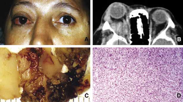
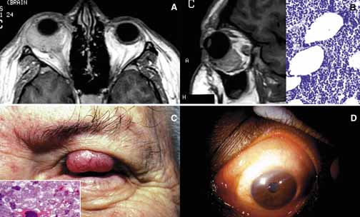
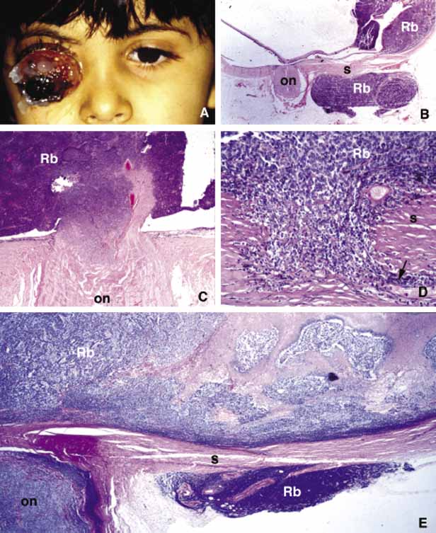
1. Ozanics V, Jakobiec F: Prenatal development of the eye and its adnexa. In Jakobiec. Ocular anatomy, embryology and teratology. Philadelphia: Harper & Row, 1982:11–96

2. Barishak YR: Embyology of the eye and its adnexa. Karger: Basel, Switzerland, 2001:15

3. Barkovich AJ: Pediatric neuroimaging, 3rd edition. New York: Raven, 2000

4. Cohen MM Jr : Craniosynostosis update. Am J Med Genet Suppl 4:99–148, 1987

5. Chumas PD, Cinalli G, Arnaud E, et al: Classification of previously unclassified cases of craniosynostosis. J Neurosurg 86:177–181, 1997

6. Casper DS, Chi TL, Trokel SL, (eds): Orbital disease, imaging and analysis. Stuttgart: Thieme, 1993

7. Beard C: Congenital hereditary abnormalities of the eyelids, lacrimal system and orbit. Transcript of New Orleans Academy of Ophthlamology. St. Louis: Mosby, 1968

8. Hertle RW, Quinn GE, Katowitz JA: Ocular and adnexal findings in patients with facial microsomias. Ophthalmology 99:114–119, 1992

9. Barkovich AJ: Neuroimaging manifestations and classification of congenital muscular dystrophies. AJNR Am J Neuroradiol 19:1389–1396, 1998

10. Waugh MC, Chong WK, Sonksen P: Neuroimaging in children with congenital disorders of the peripheral visual system. Dev Med Child Neurol 40:812–819, 1998

11. DePotter P, Shields JA, Shields CL: MRI of the eye and orbit. Philadelphia: Lippincott, 1995

12. Suwanwela C, Suwanwela N: A morphological classification of sincipital encephalomeningoceles. J Neurosurg 36:201, 1972

13. Newman NJ, Miller NR, Green WR: Ectopic brain in the orbit. Ophthalmology 93:268, 1986

14. Grossniklaus HE, Wojno T: Orbital aberrant fibroglial tissue associated with microphthalmos. J Ped Ophthalmol Strabismus 31:338–340, 1994

15. Brown HH, Kersten RC, Kulwin DR: Lipomatous hamartoma of the orbit. Arch Ophthalmol 109:240, 1991

16. Nicholson DH, Green WR: Pediatric Ocular Tumors. New York: Masson Publishing, 1981

17. Shields JA, Augsburger JJ, Donoso LA: Orbital dermoid cyst of conjunctival origin. Am J Ophthalmol 101:726, 1986

18. Smirniotopoulos JG, Chiechi MV: Teratomas, dermoids and epidermoids of the head and neck. Radiographs 15:1437–1455, 1995

19. Rochels R, Nover A, Hackelbusch R: Echographische Befunde und Differential diagnostik bei (peri-orbitalen Dermoidzyste). (Echographic findings and differential diagnosis in (peri-orbital dermoid cysts) Klin Monatsbl Augenheilkd 188:101–104,l 1986

20. Sathananthan N, Moseley IF, Rose GE, et al: the frequency and clinical significance of bone involvement in outer canthus dermoid cysts. Br J Ophthalmol 77:789–794, 1993

21. Kivela T, Tarkkanen A: Orbital germ cell tumors revisited: a clinicopathological approach to classification. Surv Ophthalmol 38:541–554, 1994

22. Lacey NA, McWilliams S, Jan W, et al: Case of the month: congenital unilateral proptosis. Br J Radiol 75:191–192, 2002

23. Soares EJC, Lopes KDS, Andrade JDG, et al: Orbital malignant teratoma. A case report. Orbit 2:235–242, 1983

24. Itani K, Traboulsi EI, Karim FWA, et al: Conservative surgery in orbital teratoma. Orbit 5:61–65, 1986

25. Hötte HH: Orbital fractures. Springfield: Charles Thomas, 1970

26. Green RP, Peters DR, et al: Force necessary to fracture the orbital floor. Ophthalmic Plast Reconst Surg 6:211, 1990

27. Fujino T, Sato TB: Mechanism of orbital blow-out fracture: experimental study by three-dimensional eye model. Orbit 6:237, 1987

28. Karcioglu ZA, Nasr AM: Diagnosis and management of orbital inflammation and infections secondary to foreign bodies: a clinical review. Orbit 17:247–267, 1998

29. Nasr AM, Haik BG, Fleming JC, Hussain H, Karcioglu ZA: Penetrating orbital injury with organic foreign bodies: a clinical review. Ophthalmology 106:523–532, 1999

30. Wojno TH: The incidence of extraocular muscle and cranial nerve palsy in orbital floor blow-out fractures. Ophthalmology 94:682, 1987

31. Ehlinger P, Peeters L, Kockx M, et al: Hematic cyst of the orbit. Acta Stomatol Belg 90:177–179, 1993

32. Law FW: Spontaneous orbital hemorrhage. Br J Ophthalmol 55:556–558, 1971

33. Cameron JD, Letson RD, Summers CG: Clinical significance of hematic cysts of the orbit. Ophthalmic Plast Reconst Surg 4:95–99, 1988

34. Shapiro A, Tso MOM, Putterman AM, et al: A clinicopathologic study of hematic cysts of the orbit. Am J Ophthalmol 102:237–241, 1986

35. Wolter JR, Fralick FB, Tanton JH: Late results of orbital hemorrhage simulating orbital neoplasm: a false aneurysm and a blood cyst with foreign body granuloma of the orbit. Am J Ophthalmol 62:528–532, 1966

36. Goldberg SH, Sassani JW, Parnes RE: Traumatic intraconal hematic cysts of the orbit. Arch Ophthalmol 110:378–380, 1992

37. Parke DW, Font RL, Boniuk M, et al: Cholesteatoma of the orbit. Arch Ophthalmol 100:612–616, 1982

38. Hill CA, Moseley IF: Imaging of orbital frontal cholesterol granuloma. Clin Radiology 46:237–242, 1992

39. Dobben GD, Fillet B, Mafee MF, et al: Orbital subperiosteal hematoma, cholesterol granuloma and infection: evaluation with MR imaging and CT. Radiol Clin North Am 36:1185–1200, 1998

40. Seabold JE, Simonson TM, Weber PC, et al: Cranial osteomyelitis: diagnosis and followup with In-111 white blood cell and Tc-99 m methylene diphosphonate bone SPECT, CT and MR imaging. Radiology 196:779–788, 1995

41. Ormerod LD, et al: Ophthalmic manifestations of maxillary sinus mucoceles. Ophthalmology 94:1013, 1987

42. Friedman DP, Rao VM, Flanders AE: Lesions causing a mass in the medial canthus of the orbit: CT and MR features. Am J Roentgenol 160:1095–1099, 1993

43. Hasso AN, Lambert D: Magnetic Resonance imaging of the paranasal sinuses and nasal cavities. Top Magn Reson Imaging 6:209–223, 1994

44. DeSouza CE, et al: Mucoceles, proptosis, and transorbital frontoethmoidectomy. Orbit 7:167, 1988

45. Sagerman RH, Chung CT, Alberti WE: Radiosensitivity of ocular and orbital structures. In Albert WE, Sagerman RH (eds): Radiotherapy of intraocular and orbital tumors. Berlin: Springer-Verlag, 1993:375–385

46. Reim M, Kuckelkom R: Chemical and thermal lesions in the orbital region. Bull Soc Belge Ophthalmol 245:21–28, 1992

47. Ervin-Mulvey LD, Nelson LB, Freeley DA: Pediatric eye trauma. Pediatr Clin North Am 30:1167–1183, 1983

48. Leonard TJK, Moseley IF, Sanders MD: Ophthalmoplegia in carotid cavernous sinus fistulas. Br J Ophthalmol 68:128–134, 1984

49. Coskun O, Hamon M, Catrous G, et al: Carotid-cavernous fistulas: diagnosis with spiral CT angiography. ANJR Am J Neuroradiol 21:712–716, 2000

50. Howard GM, Jakobiec FA, Michelsen WJ: Orbital arteriovenous malformation with secondary capillary angiomatosis treated by embolization with silastic liquid. Ophthalmology 90:1136–1139, 1983

51. Keltner JL, Satterfield D, Dublin AB, et al: Dural and carotid cavernous sinus fistulas. Ophthalmology 94:1585–1600, 1987

52. Lieb WE, Muller-Forell WS, Wichmann W: Ophthalmologic imaging methods. In, Muller-Forell WS (ed): Imaging of orbital and visual pathway pathology. Berlin: Springer 2002:9–10

53. Lieb WE, Merton DA, Shields JA: Color Doppler imaging in the demonstration of an orbital varix. Br J Ophthalmol 74:305–308, 1990

54. Bilaniuk LT: Orbital vascular lesions. Role of imaging. Radiol Clin North Am 37:169–183, 1999

55. Ambati BK, Ambati J, Azar N, et al: Periorbital and orbital cellulitis before and after the advent of Haemophilus influenzae type B vaccination. Ophthalmology 107:1450–1453, 2000

56. Givner LB: Periorbital versus orbital cellulitis. Pediatr Infect Dis J 21:1157–1158, 2002

57. Hornblass A, Herschorn BJ, Stern K, et al: Orbital abscess. Surv Ophthalmol 29:169, 1984

58. Mohammad AEA, Al-Hussaini MK, Karoosh SS, El-Moneim MTA, Carey BJ, Karcioglu ZA: Tuberculosis of the Orbit and Lacrimal Gland. Orbit 11:199–204, 1992

59. Khalil M, Lindley S, Matouk E: Tuberculosis of the orbit. Ophthalmology 108:1624–1627, 1985

60. Al-Malki FA, Issa TM, Riley F, Karcioglu ZA: Nasolacrimal tuberculosis in a patient with conjunctivodacryocystorhinostomy. Ophthal Plast Reconst Surg 15:213–216, 1999

61. Klapper SR, Partrinely JR, Kaplan SL, Font RL: Atypical mycobacterial infection of the orbit. Ophthalmology 102:1536–1541, 1995

62. Barnes PF, Bloch AB, Davidson PT, Snider DE Jr : Tuberculosis in patients with human immunodeficiency virus infection [see comments] [Review]. N Engl J Med 324:1644–1650, 1991

63. Ferry AP, Abdebi S: Diagnosis and management of rhino-orbital cerebral mucormycosis (phycomycosis): A report of 16 personally observed cases. Ophthalmology 90:1096, 1983

64. Green WR, Font RL, Zimmerman LE: Apsergillosis of the orbit: Report of ten cases and review of the literature. Arch Ophthalmol 82:302,1969

65. Karcioglu ZA, Caldwell DR: Frozen section diagnosis in ophthalmic surgery. Surv Ophthalmol 28:323, 1984

66. Margo CE, Hames LM, Ocular syphilis. Surv Ophthalmol 37:203–220, 1992

67. Dana M, Hochman MA, Viana MAG, et al: Ocular manifestations of leprosy in a noninstitutional community in the United States. Arch Ophthalmol 112:626–629, 1994

68. Ffythe T: The continuing challenge of ocular leprosy. Br J Ophthalmol 75:123–124, 1991

69. Winward KE, Smith JL, Culbertson WW, et al: Ocular Lyme borreliosis. Am J Ophthalmol 108:651–657, 1989

70. Lesser RL, Kornmehl EW, Pachner AR, et al: Neuro-ophthalmologic manifestations of Lyme disease. Ophthalmology 97(6):699–706, 1990

71. Zangwill K, et al: Cat scratch disease in Connecticut. N Engl J Med 329:8, 1993

72. Carithers HA: Oculoglandular disease of Parinaud. Am J Dis Child 132:1195, 1978

73. Blanksma L, Slijper J: Actinomycotic dacryocystitis. Ophthalmologica 176:145–49, 1977

74. Karcioglu ZA: Actinomycosis in porous polyethylene orbital implant. Graefes Arch Clin Exp Ophthalmol 235:448–451, 1997

75. Kersten RC, Shoukrey NM, Tabarra KF: Orbital myiasis. Ophthalmology 93:1228, 1986

76. Walrath JD, Lalin SC, Leib ML: Cysticercosis isolated to the orbit. Ophthal Plast Reconstr Surg 19:243–244, 2003 PMID: 12918564.

77. Rauniyar RK, Thakur SK, Panda A: CT in the diagnosis of isolated cysticercal infestation of extraocular muscle. Clin Radiol 58:154–156, 2003

78. Betharia SM, Sharma V, Pushker N: Ultrasound findings in orbital hydatid cysts. Am J Ophthalmol 135:568–570, 2003

79. Kiratli H, Bilgic S, Ozturkmen C, et al: Intramuscular hydatid cyst of the medial rectus muscle. Am J Ophthalmol 135:98–99, 2003

80. Gokcek C, Gokcek A, Akif Bayar MN, et al: Orbital hydatid cyst: CT and MRI. Neuroradiology 39:512–515, 1997

81. Awad MAN, Ray CJ, Karcioglu ZA: Echinococcus cysts of the orbit and substernum. Am J Ophthalmol 118:676–678, 1994

82. Unsold R, Greeven G: Inflammatory diseases of the orbit. Berlin: Springer Verlag, 2000

83. Bartley GB, Fatourechi V, et al: Clinical features of Graves' ophthalmopathy in an incidence cohort. Am J Ophthalmol 121:284, 1996

84. Lucarelli MJ, Shore JW: Management of thyroid optic neuropathy. International Ophth Clinics 36:179, 1996

85. Bartley GB, Gorman CA: Diagnostic criteria for Graves' ophthalmolopathy. Am J Ophthalmol 119:792, 1995

86. Leone CR, Lloyd WC: Treatment protocol for orbital inflammatory disease. Ophthalmology 92:1325, 1985

87. Kennerdell JS, Dresner SC: The nonspecific orbital inflammatory syndromes. Surv Ophthalmol 29:93–103, 1984

88. McGovern FH: The Tolusa-Hunt syndrome. Va Med 107:298–299, 1980

89. Johns ME, Wong RT: Painful ophthalmoplegia. The Tolusa-Hunt syndrome. Arch Otolaryngol 104:357–358, 1978

90. Jabs DA, Johns CJ: Ocular involvement in chronic sarcoidosis. Am J Ophthalmol 102:297–301, 1986

91. Collison MJT, et al: Involvement of orbital tissues by sarcoid. Am J Ophthalmol 102:302, 1986

92. Faris RL: Sjögren's syndrome. In Gold DH, Weingeist GA (eds): The eye in systemic disease. Philadelphia: Lippincott, 1990:70–71

93. Friedlaender MH: Ocular manifestations of Sjögren's syndrome: keratoconjunctivitis sicca. Rheum Dis Clin North Am 18:591–608, 1992

94. Robin JB, Schanzlin DJ, Meisler DM, et al: Ocular involvement in Wegener's granulomatosis. Surv Oiphthalmol 30:127–140, 1985

95. Bullen CL, Liesegang TJ, McDonald TJ, et al: Ocular complications of Wegener's granulomatosis. Ophthalmology 90:279–290, 1983

96. Power WJ, Rodriguez A, et al: Disease relapse in patients with ocular manifestations of Wegener's granulomatosis. Ophthalmology 102:154, 1995

97. Utresky SH, Kennerdell JS, Gupta JP: Graves' ophthalmolopathy in childhood and adolescence. Arch Ophthalmol 98:1963, 1980

98. Bradley E: Graves' ophthalmolopathy. Current Opinion in Ophthalmology 12:347–351, 2001

99. Balazs C, Bokk A, Molnar I: Graves' ophthalmolopathy, eye muscle antibodies and HLA antigens. Exp Clin Immunogen 6:190, 1989

100. Hufnagel TJ, Hickey WF, Cobbs WH, et al: Immunohistochemical and ultrastructural studies on the exenterated orbital tissue of a patient with Graves' disease. Ophthalmology 91:1411, 1984

101. McKenzie JM: Humoral factors in the pathogenesis of Graves' disease. Physiol Rev 48:252, 1968

102. Rootman J: Diseases of the Orbit. Philadelphia: Lippincott, 1988:241–28

103. Peyster GR, Hoover E: Computerized tomography in orbital disease and neuro-ophthalmology. Chicago: Year Book Medical Publishers, 1984:97–114.

104. Yazici B, Yazici Z, Gelisken O: An unusual case: bilateral orbital varices. Acta Ophthalmol Scand 77:453–455, 1999

105. Srivastava S, Newman J: Pseudo-pseudotumor. Surv Ophthalmol 45:135–138, 2000

106. Karcioglu ZA, Haik GB: Tissue diagnosis: Orbit. In Karcioglu ZA (ed): Laboratory Diagnosis in Ophthalmology. New York: McMillan, 1987:27–29

107. Kline LB: The Tolosa-Hunt syndrome. Surv Ophthalmol 27:79–85, 1982

108. Karcioglu ZA, Aden LB, Cruz AA, et al: Orbital invasion with prolactinoma: A clinical review of 4 patients. Ophthal Plast Reconst Surg 18:64–71, 2002

109. Mesmer EP, Font RL, McCrary JA, et al: Epitheloid angiosarcoma of the orbit presenting as Tolosa-Hunt syndrome. Ophthalmology 90:1414–1421, 1983

110. Reich JM: What is sarcoidosis? Chest 124:367–371, 2003

111. Keicho N, Kitamura K, Takaku F, et al: Serum concentration of soluble interleukin-2 receptor as a sensitive parameter of disease activity in sarcoidosis. Chest 98:112, 1990

112. Thomas PD, Hunninghake GW: Current concepts of the pathogenesis of sarcoidosis. Am Rev of Resp Diseases 135:747, 1987

113. Poulter LW: Immune aspects of sarcoidosis. Postgraduate Medical Journal 64:536–543, 1988

114. Reich JM: Deciphering histoplasmosis, systemic noncaseating granuloma, and sarcoisosis chest. 113:1143, 1998

115. Wheat LJ, French ML, Wass JL: Sarcoidlike manifestations of histoplasmosis. Arch Intern Med 149:2421–2426, 1989

116. Bronson LJ, Fisher YL: Sarcoidosis of the paranasal sinuses with orbital extensions. Arch Ophthalmol 94:243–244, 1976

117. Power WJ, Neves RA, et al: The value of combined serum angiotensin-converting enzyme and gallium scan in diagnosing ocular sarcoidosis. Ophthalmology 102:2007, 1995

118. Karcioglu ZA, Brear R: Conjunctival biopsy in sarcoidosis. Am J Ophthalmol 99:68, 1985

119. Obenauf CD, Shaw HE, Sydnor CF, et al: Sarcoidosis and its ophthalmic manifestations. Am J Ophthlamol 86:648–655, 1978

120. Talal N: Sjögren's Syndrome: Historical overview and clinical spectrum of disease. Rheum Dis Clin North Am 18:507–515, 1992

121. Callen JP, Mahl CF: Oculocutaneous manifestations observed in multisystem disorders. Dermatol Clin 10:709–716, 1991

122. Alexander EL: Neurologic disease in Sjögren's syndrome: mononuclear inflammatory vasculopathy affecting central/peripheral nervous system and muscle. Rheum Dis Clin North Am 19:869–908, 1993

123. Provost TT, Watson R: Cutaneous manifestations of Sjögren's syndrome. Rheum Dis Clin North Am 18:609–616, 1992

124. Spencer WH, Zimmerman LE: Conjunctiva. In Spencer WH (ed): Ophthalmic pathology. Philadelphia: WB Saunders, 1986:109–228

125. Jakobiec FA, Font RL: Orbit. In Spencer WH (ed): Ophthalmic pathology. Philadelphia: WB Saunders, 1986:2459–2860

126. Fox RI: Sjögren syndrome. In Sullivan DA, Dartt DA, Meneray MA (eds): Lacrimal gland, tear fluid, dry eye syndromes. New York: Plenum Press, 1998:891–902

127. Rosenbaum JT, Bennett RM: Chronic anterior and posterior uveitis and primary Sjögren's syndrome. Am J Ophthalmol 104:346–352, 1987

128. Wegener F: Uber eine eigenartige rhinogene Granulomotose mit besonderer. Beteiligung des Arteriensystems und der nieren. Beitr Pathol Anat 102:36–68, 1939

129. Straatsma BR: Ocular manifestations of Wegener's granulomatosis. Am J Ophthalmol 44:789–799, 1957

130. Greenberger MH: Central retinal artery closure in Wegener's granulomatosis. Am J Ophthalmol 63:515–516, 1967

131. Howell SB, Epstein WV: Circulating immune complexes in Wegener's granulomatosis. Am J Medicine 60:259–268, 1976

132. Falk RJ, Jennette JC: Wegener's granulomatosis, systemic vasculitis, and anti-neutrophil cytoplasmic antibodies. Annu Rev Med 42:459–469, 1991

133. Fauci AS, Wolff SM: Wegener's granulomatosis: studies in 18 patients and a review of the literature. Medicine (Baltimore) 52:535–561, 1973

134. Coutu RE, Klein M, Lessell S, et al: Limited form of Wegener's granulomatosis. Eye involvement as a major sign. JAMA 233:868–871, 1975

135. Rao NV, Wehner NG, Marshall BC, Gray WR, et al: Characterization of proteinase 3 (PR3), a neutrophil serine protease. Structural and functional. J Biol Chem 266:9540–9548, 1991

136. Ludemann G, Gross W: Autoantibodies against cytoplasmic structures of neutrophil granulocytes in Wegener's granulomatosis. Clin Exp Immunol 69:350–357, 1987

137. Gans ROB, Goldschmeding R, Donker AJM, et al: Neutrophil cytoplasmic autoantibodies and Wegener's granulomatosis. Lancet 1:269–270, 1989

138. Jennette JC, Ewert BH, Falk RJ: Do antineutrophil cytoplasmic autoantibodies cause Wegener's granulomatosis and other forms of necrotizing vasculitis? Controv Clin Rheum 19:1–14, 1993

139. Egner W, Chapel HM: Titration of antibodies against neutrophil cytoplasmic antigens is useful in monitoring disease activity in systemic vasculitides. Clin Exp Immunol 82:244–249, 1990

140. Gross WL, Ludemann G, Kiefer G: Anti-cytoplasmic antibodies in Wegener's granulomatosis. Lancet 1:806, 1986

141. Soukiasian SH, Foster CS, Niles JL, et al: Diagnostic value of antineutrophil cytoplasmic antibodies in scleritis associated with Wegener's granulomatosis. Ophthalmology 99:125–132, 1992

142. Kalina PH, Lie JT, Campbell RJ, et al: Diagnostic value and limitations of orbital biopsy in Wegener's granulomatosis. Ophthalmology 99:120–124, 1992

143. Jordan DR, Addison DA: Wegener's granulomatosis. Eyelid and conjunctival manifestations as the presenting feature in two individuals. Ophthalmolgy 101:602–607, 1994

144. Devaney KO, Travis WD, Hoffman G, et al: Interpretation of head and neck biopsies in Wegener's granulomatosis. A pathologic study of 126 biopsies in 70 patients. Am J Surg Pathol 14:555–564, 1990

145. Del Buono EA, Flint A: Diagnostic usefulness of nasal biopsy in Wegener's granulomatosis. Hum Pathol 22:107–110, 1991

146. Travis WD, Hoffman GS, Leavtitt RY, et al: Surgical pathology of the lung in Wegener's granulomatosis. Review of 87 open lung biopsies from 67 patients. Am J Surg Pathol 15:315–333, 1991

147. Fauci AS, Haynes BF, Katz P, et al: Wegener's granulomatosis: prospective clinical and therapeutic experience with 85 patients for 21 years. Ann Intern Med 98:76–85, 1983

148. Koorneef L, Melief CJM, Peterse HL, et al: Wegener's granulomatosis of the orbit. Orbit 2:1–10, 1983

149. Specks U, DeRemee RA: Granulomatous vasculitis. Wegener's granulomatosis and Churg-Strauss syndrome. Rheum Clin North Am 16:377–397, 1990

150. Foster CS: Immunosuppressive therapy in external ocular inflammatory disease. Ophthalmology 87:140–150, 1980

151. Chang WJ, Shields CL, Shields JA, et al: Bilateral orbital involvement with massive allergic fungal sinusitis. Arch Ophthalmol 114:767–768, 1996

152. Dunlop IS, Billson FA: Visual failure in allergic aspergillus sinusitis: case report. Br J Ophthalmol 72:127–130, 1988

153. Torres C, El Naggar AK, Sim SJ, et al: Allergic fungal sinusitis: a clinicopathological study of 16 cases. Human Pathol 27:793–799, 1996

154. Michaels L, Lloyd G, Phelps T: Origin and spread of allergic fungal disease of the nose and paranasal sinuses. Clin Otolaryngol Allied Sci 25:518–525, 2000

155. Ordonez NG: Application of immunocytochemistry in the diagnosis of soft tissue sarcomas: a review and update. Adv Anat Pathology 5:67–85, 1998

156. Folpe AL, Gown AM: Immunohistochemistry for analysis of soft tissue tumors. In Weiss SW, Goldblum JR (eds): Soft tissues tumors, 4th edition. St. Louis: Mosby, 2001

157. Swanson SA, Brooks JJ: Proliferation markers Ki-67 and p105 in soft tissue lesions: correlation with DNA flow cytometric characteristics. Am J Pathol 137:1491–1500, 1990

158. Hoos A, Stojadonovic A, Mastorides S, et al: High Ki-67 proliferative index predicts disease specific survival in patients with high risk soft tissue sarcoma. Cancer 92:869–874, 2001

159. Karcioglu ZA, Haik BG: Tissue Diagnosis: Orbit. In Karcioglu ZA (ed): Laboratory Diagnosis in Ophthalmology. New York: MacMillan Publishing Co., 1986:25–52

160. Burnstine MA, Fruch BR, Elner VM: Angiosarcoma metastatic to the orbit. Arch Ophthalmol 114:93–96, 1996

161. Shields JA, Shields CL, Brotman HK, et al: Cancer metastatic to the orbit: the 2000 Robert M. Curts Lecture. Ophthal Plast Reconstr Surg 17:346–354, 2001

162. Char DH, Miller T, Kroll S: Orbital metastases: Diagnosis and course. Br J Ophthalmol 81:386, 1997

163. Shields JA, Bakewell B, Augsburger JJ, et al: Classification and incidence of space-occupying lesions of the orbit. Arch Ophthalmol 102:1601–1611, 1984

164. Henderson JW, Campbell RJ, Farrow GM, et al: Orbital Tumors. New York: Raven Press, 1994:43–52

165. Gunalp I, Gunduz K: Biopsy-proven orbital lesions in Turkey. A survey of 1092 cases over 30 years. Orbit 13:67–69, 1994

166. Seregard S, Sahlin S: Panaorama of orbital space-occupying lesions. The 24-year experience of a referral center. Acta Ophthalmol Scand 77:91–98, 1999

167. Jakobiec FA, Bilyk JR, Font RL: Lacrimal gland tumors. In Spencer WH (ed): Ophthalmic Pathology. An Atlas and Textbook, 4th edition. Philadelphia: WB Saunders Company, 1996:2485–2525

168. Grossniklaus HE, Abbuhl MF, McLean IW: Immunohistologic properties of benign and malignant mixed tumor of the lacrimal gland. Am J Ophthalmol 110:540–549, 1990

169. Shields JA, Shields CL: Malignant transformation of presumed pleomorphic adenoma of lacrimal gland after 60 years. Arch Ophthamol 105:1403, 1987

170. Tellad MV, McLean I, Specht CS, et al: Adenoid cystic carcinomas of the lacrimal gland in childhood and adolescence. Ophthalmology 104:1622–1625, 1997

171. Wright JE, Rose GE, Garner A: Primary malignant neoplasms of the lacrimal gland. Br J Ophthalmol 76:401–407, 1992

172. Gamel JW, Font RL: Adenoid cyst carcinoma of the lacrimal gland: the clinical significance of a basaloid histologic pattern. Hum Pathol 13:219–225, 1982

173. Eviatar JA, Hornblass A: Mucoepidermoid carcinoma of the lacrimal gland: 25 cases and a review and update of the literature. Ophthal Plast Reconstr Surg 9:170–181, 1993

174. Levin LA, Popham J, To K, et al: Mucoepidermoid carcinoma of the lacrimal gland: Report of a case with oncocytic features arising in a patient with chronic dacryops. Ophthalmology 98:1551, 1991

175. Bullock JD, Fleishman JA, Rosset JS: Lacrimal duct cysts. Ophthalmology 93:1355–1360, 1986

176. Monica M, Long DA, Karcioglu ZA: Bilateral dacryops. Ann Ophthalmol 20:259–263, 1988

177. Margo CE, Karcioglu ZA, Naugle TC: Ectopic lacrimal gland tissue of the orbit and sclerosing dacryoadenitis. Ophthalmic Surg 16:178, 1985

178. Harris GJ: Orbital vascular malformations: A consensus statement on terminology and its clinical implications. Am J Ophthalmol 127:453–455, 1999

179. Haik BG, Karcioglu ZA, Gordon RA, Pechos B: Capillary Hemangioma. [Major Review]. Survey Ophthalmol 38:399–426, 1994

180. Chang E, Boyd A, Nelson CC, et al: Successful treatment of infantile hemangiomas with interferon-alpha–2B. J Pediatr Hematol Oncol 19:237–244, 1997

181. Burstein FD, Simms C, Cohen SR, et al: Intralesinal laser therapy of extensive hemangiomas in 100 consecutive pediatric patients. Ann Plast Surg 44:188–194, 2000

182. Kushner BJ: Intralesional corticosteroid injection for infantile adnexal hemangioma. Am J Ophthalmol 93:496–506, 1982

183. Karcioglu ZA, Nasr MN, Haik BG: Orbital hemangiopericytoma: Clinical and morphologic features. Am J Ophthalmol 124:661–672, 1997

184. Rice CD, Kersten RC, Mrak RE: An orbital hemangiopericytoma recurrent after 33 years. Arch Ophthalmol 107:552, 1989

185. Harris GJ, Jakobiec FA: Cavernous hemangioma of the orbit: A clinicopathologic analysis of sixty-six cases. In Jakobiec FA (ed): Ocular and Adnexal Tumors, Birmingham AL:. Aesculapius 1978:741

186. Sullivan TJ, Aylward GW, Wright JE, et al: Bilateral multiple cavernous hemangiomas of the orbit. Br J Ophthalmol 76:627–629, 1992

187. Henderson JW, Campbell RJ, Farrow GM, et al: Orbital Tumors. New York: Raven Press, 1994:89–118

188. Tunc M, Sadri E, Char DH: Orbital lymphangioma: an analysis of 26 cases. Br J Ophthalmol 83:760–780, 1999

189. Rootman J, Hay E, Graeb D, et al: Orbital-adnexal lymphangiomas: A spectrum of hemodynamically isolated vascular hamartomas. Ophthalmology 93:1558, 1986

190. Harris GJ, Sakel PJ, Bonavolonta G, et al: An analysis of thirty cases of orbital lymphangioma: Pathophysiologic considerations and management recommendations. Ophthalmology 97:1583, 1990

191. Shields JA, Dolinskas C, Augsburger JJ, et al: Demonstration of orbital varix with computed tomography and valsalva maneuver. Am J Ophthalmol 97:108–110, 1984

192. Keltner J, Satterfield, D, Dublin AB, et al: Dural and carotid cavernous sinus fistulas. Ophthalmology 94:1585–1600, 1987

193. Wright JE, Sullivan TJ, Garner A: Orbital venous anomalies. Ophthalmology 104:905–913, 1997

194. Gunduz K, Shields JA, Shields CL, et al: Cutaneous angiosarcoma with eyelid involvement. Am J Ophthalmol 125:870–871, 1998

195. Hufnagel T, Ma L, Kuo TT: Orbital angiosarcoma with subconjunctival presentation. Ophthalmology 94:72–77, 1987

196. Shields JA, Shields CL, Eagle RC Jr , et al: Intravascular papillary endothelial hyperplasia with presumed bilateral orbital varices. Arch Ophthalmol 117:1247–1249, 1999

197. Jolly SS, Brownstein S, Jordan DR: Leiomyoma of the anterior orbit and eyelids. Can J Ophthalmol 30:366–370, 1995

198. Kaulkarni V, Rajshekhar V, Chandi SM: Orbital apex leiomyoma with intracranial extension. Surg Neurol 54:327–330, 2000

199. Smith DL, Kincaid MC, Nicolitz E: Angiolymphoid hyperplasia with eosinophilia (Kumura's disease) of the orbit. Arch Ophthalmol 106:793–795, 1988

200. Karcioglu ZA, Yulug A, Haik BG: Tumors of the optic nerve. In, Margo C, et al (eds): Diagnostic Problems in Clinical Ophthalmology. Philadelphia: Saunders, 1994:105–106

201. Alvord EJ, Lofton S: Gliomas of the optic nerve or chiasm. Outcome by patient's age, tumor site and treatment. J Neurosurg 68:85–98, 1988

202. Dutton JJ: Gliomas of the anterior visual pathway. Surv Ophthalmol 38:427–452, 1994

203. Brodovsky S, Hove MW, Pinkerton RM, et al: An enhancing optic nerve lesions: malignant glioma of adulthood. Can J Ophthalmol 32:409–413, 1997

204. Chateil JF, Sousotte C, Pedespan JM, et al: MRI and clinical differences between optic pathway tumours in children with and without neurofibromatosis. Br J Radiology 74:24–31, 2001

205. Deliganis AV, Geyer JR, Berger MS: Prognostic significance of type I neurofibromatosis (von Recklinghausen Disease) in childhood optic glioma. Neurosurgery 38:1114–1118, 1996

206. Lee AG, Dutton J: A practice pathway for the management of gliomas of the anterior visual pathway: an update and an evidence-based approach. Neuroophthalmology 22:139–155, 1999

207. Levin LA, Jakobiec FA: Optic nerve tumors of childhood: A decision analytical approach to their diagnosis. Int Ophthalmol Clin 32:223–240, 1992

208. Mulvihill JJ, Parry MD, Sherman JL, et al: NIH Conference. Neurofibromatosis 1 (Recklinghausen disease) and neurofibromatosis 2 (bilateral acoustic neurofibromatosis. An update. Ann Intern Med 113:39–52, 1990

209. Pearson-Webb MA, Kaiser-Kupfer MI, Eldridge R: Eye findings in bilateral acoustic (central) neurofibromatosis: Association with presenile lens opacities and cataracts but absence of Lisch nodules (letter). N Engl J Med 35:1553–1554, 1986

210. Cummings TJ, Provenzale JM, Hunter SB, et al: Gliomas of the optic nerve: histological, immunohistochemical (MIB–1 and p53), and MRI analysis. Acta Neuropathologica 99:563–570, 2000

211. Bilgic S, Erbengi A, Tinaztepe B, et al: Optic glioma of childhood: Clinical, histopathological, and histochemical observations. Br J Ophthalmol 73:832–837, 1989

212. Imes RK, Hoyt WF: Magnetic resonance imaging signs of optic nerve gliomas in neurofibromatosis. Am J Ophthalmol 111:729–734, 1991

213. Stern J, Jakobiec FA, Housepian EM: The architecture of optic nerve gliomas with and without neurofibromatosis. Arch Ophthalmol 98:505–511, 1980

214. Cooling RJ, Wright JE: Arachnoid hyperplasia in optic nerve glioma: Confusion with orbital meningioma. Br J Ophthalmol 63:596–599, 1979

215. Fuss M, Hug EB, Schaefer RA, et al: Proton radiation therapy (PRT) for pediatric optic pathway gliomas: comparison with 3D planned conventional photons and standard photon technique. Int J Rad Oncol Biol Physics 45:1117–1126, 1999

216. Muci-Mendoza R, Arevalo JF, Ramella M, et al: Optociliary veins in optic nerve sheath meningioma. Indocynanine green videoangiography findings. Ophthalmology 106:311–318, 1999

217. Cunliffe IA, Moffat DA, Hardy DG, et al: Bilateral optic nerve sheath meningiomas in a patient with neurofibromatosis type 2. Br J Ophthalmol 76:310–312, 1992

218. Mafee MF, Goodwin J, Dorodi S: Optic nerve sheath miningiomas. Role of MR imaging. Radiol Clin North Am 37:37–58, 1999

219. Turbin RE, Thompson CR, Kennderdell JS, et al: A long-term visual outcome comparison in patients with optic nerve sheath meningioma managed with observation, surgery, radiotherapy, or surgery and radiotherapy. Ophthalology 109:890–900, 2002

220. Moyer PD, Golnik KC, Breneman J: Treatment of optic nerve sheath meningioma with three-dimensional conformal radiation. Am J Ophthalmol 129:694–696, 2000

221. Rootman J, Goldberg C, Robertson W: Primary orbital Schwannomas. Br J Ophthalmol 66:194–204, 1982

222. Carroll GS, Haik BG, Fleming JC, et al: Peripheral nerve tumors of the orbit. Rad Clin North Am 37:195–202, 1999

223. Rose GE, Wright JE: Isolated peripheral nerve sheath tumours of the orbit. Eye 5:668–673, 1991

224. Abet T, Kawamura N, Homma H, et al: MRI of orbital schwannomas. Head Neck Radiol 42:466–468, 2000

225. Schatz H: Benign orbital neurilemmoma: sarcomatous transformation in von Recklinghausen's disease. Arch Ophthalmol 86:268–273, 1971

226. Harkin JC, Reed RJ: Tumors of the peripheral nervous system. Atlas of tumor pathology. Washington, DC, 1968:29–59

227. Lam DSC, Ng JSK, To TAF, et al: Cystic schwannoma of the orbit. Eye 11:798–800, 1997

228. Jakobiec FA, Font RL, Zimmerman LE: Malignant peripheral nerve sheath tumors of the orbit: a clinical pathological study of eight cases. Trans Am Ophthalmol Soc 83:332–366, 1985

229. Lewis RA, Gerson LP, Axelson KA, et al: Von Recklinghausen neurofibromatosis. II. Incidence of optic gliomata. Ophthalmology 91:929–935, 1984

230. Henderson JW, Campbell RJ, Farrow GM, et al: Orbital Tumors. New York: Raven Press, 1994:221–37

231. Jackson IT, Laws ERJr , Martin RD: The surgical management of orbital neurofibromatosis. Plast Reconst Surg 71:751–758, 1983

232. Bednar MM, Trainer TD, Aitken, PA, et al: Orbital paraganglioma: case report and review of the literature. Br J Ophthalmol 76:183–185, 1992

233. Gunalp I, Gunduz K, Duruk K, Kanpolat Y: Neurogenic tumors of the orbit. Jpn J Ophthalmol 38:185–190, 1994

234. Myssiorek D: Head and neck paragangliomas: an overview. Otolaryngol Clin North Am 34:829–836, 2001

235. Castillo BVJr , Kaufman L: Pediatric tumors of the eye and orbit. Pediatr Clin North Am 50:149–172, 2003

236. He Y, Song G, Ding Y: Histopathologic classification of 3,476 orbital diseases. Zhonghua Yan Ke Za Zhi 38:396–398, 2002

237. Shields JA, Shields CL: Rhabdomyosarcoma: review for the ophthalmologist. Surv Ophthalmol 48:39–57, 2003

238. Knowles DM II , Jakobiec FA, Potter GD, et al: Ophthalmic striated muscle neoplasms. Surv Ophthalmol 21:219–261, 1976

239. Margo CE, Karcioglu ZA: Diagnostic electronmicroscopy In Karcioglu ZA (ed): Laboratory Diagnosis in Ophthalmology. New York: McMillian, 1987:120–130

240. Buwalda J, Schouwenburg PF, Blank LE, et al: A novel local treatment strategy for advanced stage head and neck rhabdomyosarcomas in children: results of the AMORE protocol. Eur J Cancer 39:1594–602, 2003

241. Seregard S: Management of alveolar rhabdomyosarcoma of the orbit. Acta Ophthalmol Scand 80:660–664, 2002

242. Luckenbach M: Idiopathic inflammatory pseudotumor of childhood. Park City, UT:. Hogan-Theobald Society Meeting, 1984

243. Shields CL, Shields JA, Honavar SG, Demirci H: Primary ophthalmic rhabdomyosarcoma in 33 patients. Trans Am Ophthalmol Soc 99:133–142, 2001

244. Fetkenhour DR, Shields CL, Chao , et al: Orbital cavitary rhabdomyosarcoma masquerading as lymphangioma. Arch Ophthalmol 11:1208–1210, 2001

245. Seedat RY, Hamilton PD, de Jager LP, et al: Orbital rhabdomyosarcoma presenting as an apparent orbital subperiosteal abscess. Int J Pediatr Otorhinolaryngol 52:177–181, 2000

246. Gordon LK: Diagnostic dilemmas in orbital inflammatory disease. Ocul Immunol Inflamm 11:3–15, 2003

247. Hou LC, Murphy MA, Tung GA: Primary orbital leiomysarcoma: a case report with MRI findings. Am J Ophthalmol 135:408–410, 2003

248. Klippenstein KA, Wesley RE, Glick AD: Orbital leiomyosarcoma after retinoblastoma. Ophthalmic Surg Lasers 30:579–583, 1999

249. Weichens B, Werner JA, Luttges J, et al: Primary orbital leimyoma and leiomyosarcoma. Ophthalmologica 213:159–164, 1999

250. Karcioglu ZA, Carroll G, Nasr A, Haik BG: Fibrous lesions of the orbit and periorbital tissues. Amsterdam: XXVIII International Congress of Ophthalmology (ICO), 1998:8

251. Dalley RW: Fibrous histiocytoma and fibrous tissue tumors of the orbit. Radiol Clin North Am 1999;37:185–194

252. Jakobiec FA, Font RL: Orbit. In Spencer WH, Font RL, Green WR, et al (eds): Ophthalmic Pathology. An Atlas and Textbook. Vol. 3. Philadelphia: W.B. Saunders Co., 1986:2568–2580

253. Caballero LR, Rodriguez AC, Sopelana AB: Angiomatoid fibrous histiocytoma of the orbit. Am J Ophthalmol 92:13–15, 1981

254. Lui D, McCann P, Kini RK, et al: Malignant fibrous histiocytoma of the orbit in a 3-year-old girl. Arch Ophthalmol 105:895–896, 1987

255. Font RL, Hidayat AA: Fibrous histiocytoma of the orbit. A clinicopathologic study of 150 cases. Hum Pathol 13:199–209, 1982

256. Ros PR, Kursunoglu S, Batlle JF, et al: Malignant fibrous histiocytoma of the orbit. J Clin Neuro-ophthalmol 5:116–119, 1985

257. Masson JK, Soule EH: Desmoid tumors of the head and neck. Am J Surg 112:6115–6122, 1966

258. Perry RH, Ramani PS, McAllister V, et al: Nodular fasciitis causing unilateral proptosis. Br J Ophthalmol 59:404–408, 1975

259. Font RL, Zimmerman LE: Nodular fasciitis of the eye and adnexa. A report of ten cases. Arch Ophthalmol 75:475–481, 1966

260. Ferlito A, Recher G, Tomazzolli L: Radiation-induced fibrosarcoma of the mandible following treatment for bilateral retinoblastoma. J Laryngol Otol 93:1015–1020, 1979

261. Karcioglu ZA, Al-Rasheed W, Gray A: Second neoplasms in retinoblastoma. Middle East J Ophthalmol 5:99–104, 1997

262. Polito E, Tosi M, Toti P, et al: Orbital solitary fibrous tumor with aggressive behavior. Three cases and the review of the literature. Grafes Arch Exp Ophthalmol 240:570–574, 2002

263. Kim HY, Lee SY, Kang SJ, et al: Solitary fibrous tumor of the orbit: a poorly recognized orbital lesion. Acta Ophthalmol Scand 77:704–708, 1999

264. Lanuza A, Lazaro R, Salvador M, et al: Solitary fibrous tumour of the orbit. Report of a new case. Int Ophthalmol 22:265–268, 1998

265. Caroll G, Haik BG, Karcioglu ZA: Orbital giant cell fibroblastoma. Orbit 18:25–32, 1999

266. Van Furth R, Cohn ZA, Hirsch JG, et al: The mononuclear phagocyte system: a new classification of macrophages, monocytes and their precursor cells. Bull World Health Org 46:845–855, 1972

267. Chu AC: The confusion state of the histicytoses. Br J Dermatol 143:475–476, 2000

268. Karcioglu ZA, Sharara N, Boles, TL, Nasr AM: Orbital xanthogranuloma, clinical and morphologic features in 8 patients. Ophthal Plast Reconstr Surg 19:372–381, 2003

269. Malone M: The histiocytosis of childhood. Histopathology 19:105–119, 1991

270. Dahlmann AH, Simmons I, Picton S, et al: Single-Langerhans cell histiocytosis of the orbit. Eye 14:109–110, 2000

271. Veyssier-Belot C, Cacoub P, Caparros-Lefebvre D, et al: Erdheim-Chester disease. Clinical and radiologic characteristics of 59 cases. Medicine (Baltimore) 75:157–169, 1996

272. Chetritt J, Paradis V, Dargere D, et al: Chester-Erdheim disease: a neoplastic disorder. Hum Pathol 30:1093–1096, 1999

273. Alper MG, Zimmerman LE, la Piana FG: Orbital manifestations of Erdheim-Chester disease. Trans Am Ophthalmol Soc 81:64–85, 1983

274. Shields JA, Karcioglu ZA, Shields CL, et al: Orbital and eyelid involvement with Erdheim-Chester disease. A report of two cases. Arch Ophthalmol 109:850–854, 1991

275. Miszkiel KA, Sohaib SAA, Rose GE: Radiological and clinicopathological feaures of orbital XG. Br J Ophthalmol 84:251–258, 2000

276. Karcioglu ZA: Bone and cartilage tumors. In Karcioglu ZA (ed): Orbital Tumors. New York: Springer-Verlag, 2004:(in press)

277. Earwaker J: Perinasal sinus osteomas: a review of 46 cases. Skel Radiol 22:417–423, 1993

278. Tucker MA, D'Angio GJ, Boice JD Jr. , et al: Bone sarcomas linked to radiotherapy and chemotherapy in children. N Engl J Med 317:588–593, 1987

279. Shields JA: Lymphoid tumors and leukemias. In Shields JA (ed): Diagnosis and Management of Orbital Tumors, Philadelpha: WB Saunders, 1989:316

280. Jakobiec FA, Font RL: Orbit: lymphoid tumors. In Spencer WH, Font RL, Green WR, et al (eds): Ophthalmic Pathology: An Atlas and Textbook. Vol 3, ed. Philadelphia: WB Saunders Co., 1986:2663–2711

281. Sharara N, Holden JT, Wojno TH, et al: Ocular adnexal lymphoid proliferations. Clinical, histological, flow cytometry and molecular analysis of 43 cases. Ophthalmology 110:1245–1254, 2003

282. Freedman AS, Nadler LM: NonHodgkin's lymphomas. In Bast RC, Kufe DW, Pollock RE, et al (eds): Cancer Medicine 5th ed. BC Decker Inc., 2000:Section 36, Ch 130

283. Chan JKC: Tumors of the lymphoreticular system, including skin and thymus. In, Fletcher CDM (ed): Diagnostic histopathology of tumors, 2nd ed. London: Churchill Livingston, 2000:1099–1316

284. Harris NI, Jaffe ES, Stein H, et al: A revised European-American classification of lymphoid neoplasms: a proposal from the International Lymphoma Study Group. Blood 84:1361–1392, 1994

285. Armitage JO, Weisenburger DD: New approach to classifying nonHodgkin's lymphomas: Clinical features of the major histologic subtypes. J Clin Oncol 16:178–195, 1998

286. Coupland SE, Krause L, Des Velecluse HJ, et al: Lymphoproliferative lesions of the ocular adnexa: analysis of 112 cases. Ophthalmology 105:1430–1441, 1998

287. Jenkins C, Rose GE, Bunce C, et al: Histological features of ocular adnexal lymphoma (REAL classification) and their association with patient morbidity and survival. Br J Ophthalmol 84:907–913, 2000

288. Jakobiec FA, Lefkowich J, Knowles DM: B- and T-lymphocytes in ocular disease. Ophthalmology 91:635, 1984

289. Coupland SE, Hummel M, Stein H: Ocular adnexal lymphomas, five case presentations and a review of the literature. Surv Ophthalmol 47:470–490, 2002

290. Au Eong, Choo CT: Burkitt lymphoma manifesting as acute proptosis. Am J Ophthalmol 123:856–858, 1997

291. Uceda-Montanes A, Blanco G, Saornil MA, et al: Extramedullary plasmocytoma of the orbit. Acta Ophthalmol Scand 78:601–603, 2000

292. Rodman HI, Font RL: Orbital involvement in multiple myeloma: Review of the literature and report of three cases. Arch Ophthalmol 87:30, 1972

293. Kennedy RE: An evaluation of 820 orbital cases. Trans Am Ophthalmol Soc 82:134, 1984

294. Rootman J: Disease of the orbit. A multidisciplinary approach. Philadelphia: JB Lippincott Co., 1988:405–427

295. Jensen OA: Metastatic tumors of the eye and orbit: a histopathologic analysis of a Danish series. Acta Pathol Microbiol Scand 212:201, 1970

296. Font RL, Ferry AP: Carcinoma metastatic to the eye and orbit. III: A clinicopathologic study of 28 cases metastatic to the orbit. Cancer 38:1326, 1976

297. Shields CL, Shields JA, Peggs M: Tumors metastatic to the orbit. Ophthalmic Plast Reconstr Surg 4:73, 1988

298. Henderson JW, Campbell RJ, Farrow GM, Garrity JA: Orbital tumors, 3rd ed. New York: Raven Press, 1993;361–375

299. Shields JA, Shields CL, Brotman HK, et al: Cancer metastatic to the orbit: the 2000 Robert M. Curts Lecture. Ophthal Plast Reconstr Surg 17:346–354, 2001

300. Goldberg RA, Rootman J, Cline RA: Tumors metastatic to the orbit: A changing picture. Surv Ophthalmol 35:1, 1990

301. Katz SE, Rootman J, Goldberg RA: Secondary and metastatic tumors of the orbit. In Tasman W, Jaeger EA (eds): Duane Ophthalmology CD-ROM Edition. Hagerstown, MD:. Lippincott, Williams & Wilkins, 2001:Clinical Volume 2, Chapter 46

302. Healy JF: Computed tomographic evaluation of metastases to the orbit. Ann Ophthalmol 15:1026, 1983

303. Peyster RG, Shapiro MD, Haik BG: Orbital metastasis: Role of magnetic resonance imaging and computed tomography. Radiol Clin North Am 25:647, 1987

304. Dresner SD, Kennerdell JS, Dekker A: Fine needle aspiration biopsy of metastatic orbital tumors. Surv Ophthalmol 27:397, 1983

305. Kennerdell JS, Slamovits TL, Dekker A, et al: Orbital fine needle aspiration biopsy. Am J Ophthalmol 99:547, 1985

306. Das DK, Das J, Bhatt NC, et al: Orbital lesions. Diagnosis by fine needle aspiration cytology. Acta Cytol 38:158–164, 1994

307. Krohel GB, Tobin DR, Chavis RM: Inaccuracy of fine needle aspiration biopsy. Ophthalmology 92:666, 1985

308. Liu D: Complications of fine needle aspiration biopsy of the orbit. Ophthalmology 92:1768, 1985

309. Shetlar DJ, Font RL, Ordonez N, et al: A clinicopathologic study of three carcinoid tumors metastatic to the orbit. Ophthalmology 97:257, 1990

310. Fan JT, Buettner H, Bartley GB, Bolling JP: Clinical features and treatment of seven patients with carcinoid tumor metastatic to the eye and orbit.Am J Ophthalmol 119:211, 1995

311. Amemiya T, Hayashida H, Dake Y: Metastatic orbital tumors in Japan: a review of the literature. Ophthalmic Epidemiol 9:35–47, 2002

312. Evans AR: Congenital neuroblastoma. J Clin Pathol 18:54–62, 1965

313. Reynolds P, Smith R, Frenkel B: The diagnostic dilemma of small, round cell neoplasm. Cancer 48:2088, 1981

314. Henderson JW, Farrow GM: Orbital tumors (2nd ed.), New York: Brian C. Decker (Thieme-Stratton), 1980:425–450

315. Crotty TB, Scheithauer BW, Young WF, et al: Papillary craniopharyngioma: a clinical study of 48 cases. J Neurosurg 83:206–214, 1995

316. Wilson WB: Meningiomas of the anterior visual system. Surv Ophthalmol 26:109, 1981

317. Carrau RL, Segas J, Nuss DW, et al: Squamous cell carcinoma of the sinonasal tract invading the orbit. Laryngoscope 109:230–235, 1999

318. Karcioglu ZA, et al: Proptosis pseudocyst formation from inverted papilloma. Ann Ophthalmol 14:443–448, 1982

319. Hurwitz JJ: Lacrimal sac tumours. Chapter 28, In Hurwitz JJ (ed): The Lacrimal System. Philadelphia: Lippincott-Raven, 1996:195

320. Shields JA, Shields CL: Orbital malignant melanoma: 2002 Sean B. Murphy lecture. Ophthalmol Plas Reconstr Surg 19:262–269, 2003

321. Finger PT, Harbour JW, Karcioglu ZA: Risk factors for metastasis in retinoblastoma. Surv Ophthalmol 47:1–16, 2002

322. Liesegang GK: Amyloid infiltration of the levator palpebral superioris muscle. Ann Ophthalmol 15:610–613, 1983

323. Gorevic PD, Rodrigues MM: Ocular amyloidosis. Am J Ophthalmol 113:529–532, 1994

324. Sevino PJ, Schatz NJ, Rodrigues MM: Orbital amyloidosis. Can J Ophthalmol 11:252–255, 1976

325. Wheeler GE, Eiferman RA: Immunohistochemical identification of the AA protein in lattice dystrophy. Exp Eye Research 36:181, 1983

326. Breathnach SN: Amyloid and amyloidosis. J Am Acad Derm 18:1–16, 1988

327. Okamoto K, Ito J, Emura T, et al: Focal orbital amyloidosis presenting as rectus muscle enlargement: CT and MR findings. AJNR Am J Neuroradiol 19:1799–1801, 1988

328. Murdoch IE, Sullivan TJ, Moseley I, et al: Primary localized amyloidosis of the orbit. Br J Ophthalmol 80:1083–1086, 1996

329. Massry GG, Harrison W, Hornblass A: Clinical and computed tomographic characteristics of amyloid tumors of the lacrimal gland. Ophthalmology 103:1233–1236, 1996

330. Ustun MO, Ekinci N, Payzin B: Extramedullary plasmocytoma of the parotid gland. Report of a case with extensive amyloid deposition masking the cytology and histopathologic picture. Acta Cytol 45:449–453, 2001

331. Jabs DA, Miller NA, Neuman SA, et al: Optic neuropathy in systemic Lupus erythematosus. Arch Ophthalmol 104:564, 1986

332. Grimson BS, Simons KB: Orbital inflammation, myositis, and systemic lupus erythematosis. Arch Ophthalmol 101:736–738, 1983

333. Ibanez HE, Bardenstein DS, Korman NJ, et al: Exuberant conjunctival pseudopolyposis in a patient with dermatomyositis. Ann Ophthalmol 25:326–327, 1993

334. Leib ML, Odel JG, Cooney MJ: Orbital polymyositis and giant cell myocarditis. Ophthalmology 101:950–954, 1994

335. Selva-O'Callaghan A, Mijares-Boeckh-Behrens T, Solans-Laque R, et al: Dermatomyositis and Graves' disease. Clin Exp Rheumatol 19:595–596, 2001

336. Watson PG, Hazelman BL: The sclera and systemic disorders. Philadelphia: WB Saunders, 1976

337. Foster CS, Sainz de la Masa M: The sclera. New York: Springer-Verlag, 1994

338. Horan EC: Ophthalmic manifestations of progressive systemic sclerosis. Br J Ophthalmol 53:388–392, 1969

339. Manschot WA: Generalized scleroderma with ocular symptoms. Ophthalmologica 149:131–137, 1965

340. Anzai H, Tajima S: Systemic scleroderma associated with Graves' disease. J Dermatol 23:896–898, 1996

341. Nicholson D, White S, Lipson A, et al: Progressive systemic sclerosis and Graves' disease. Report of three cases. Arch Intern Med 146:2350–2352, 1986

342. Koike R, Yamada M, Matsunaga T, et al: Polyarteritis nodosa (PN) complicated with unilateral exophthalmos. Intern Med 32:232–236, 1993

343. Ishida K, Yokota T, Wada Y, et al: Unilateral facial swelling and exophthalmos in a patient with polyarteritis nodosa. Intern Med 31:500–503, 1992

344. Prasad TS, Suresh PS, Rodrigues G, et al: Orbital involvement with angiolymphois hyperplasia eosinophilia—a benign condition for the practicing ophthalmologist to be aware of. Eye 14:390–392, 2000

345. Buggage RR, Spraul CW, Wojno TH, et al: Kimura disease of the orbit and ocular adnexa. Surv Ophthalmol 44:79–91, 1999

346. Cook HT, Stafford ND: Angiolymphoid hyperplasia with eosinophilia involving the lacrimal gland: case report. Br J Ophthalmol 72:710–712, 1988

347. Francis IC, Kappagoda MB, Smith J, et al: Kimura's disease of the orbit. Ophthal Plast Reconstr Surg 4:235–239, 1988

348. Al-Malki AF, Hussain H, Al-Ma'ani JR, et al: Extraocular myositis in Behcet's Syndrome. VI International Symposium on Ocular Inflammation. Istanbul, Turkey, June 2000

349. Sale S, Patterson R: Recurrent Churg-Strauss vasculitis. With exophthalmos, hearing loss, nasal obstruction, amyloid deposits, hyperimmunoglobulinemia E, and circulating immune complexes. Arch Intern Med 141:1363–1365, 1981

350. Iliff NT: The ophthalmic implications of the correction of late enophthalmos following severe midfacial trauma. Trans Am Ophthalmol Soc 89:455–548, 1991

351. Cline RA, Rootman J: Enophthalmos. A clinical review. Ophthalmology 91:229, 1984

352. Nasr AM, Ayyash I, Karcioglu ZA: Unilateral enophthalmos secondary to acquired hemilipodystrophy. Am J Ophthalmol 124:572–575, 1997

Back to Top