|
|
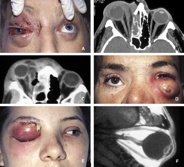
| Fig. 6 Orbital cellulitis. A and B show the appearance of right orbital cellulitis secondary to Staphylococcus infection from right ethmoidal sinus. The proptosis, chemosis and visual loss in this patient worsened after the ethmoidectomy and the medial orbit had to be explored with additional drainage of pus. Note the stretch of the right optic nerve and pear-shaped right globe secondary to marked proptosis on an axial CT scan (B). C shows another case with severe ethmoiditis causing cellulitis of the right orbit with the extension of the infection preseptally toward the left globe. The patient responded well to ethmoidectomy and drainage of the abscess from the right medial orbit. D shows the appearance of a preseptal and anterior orbital cellulitis secondary to a long-standing foreign body caused in a motor vehicle accident. The patient depicted in frame E developed Streptococcal orbital cellulitis with secondary brain abscess as depicted in the sagittal T1-weighted MRI (F). |