|
|
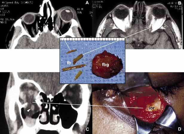
| Fig. 3 Orbital trauma. Axial CT scan (A) and T1-weighted MRI (B) show multiple organic foreign bodies (fb) within the left medial rectus muscle and posterior orbit. The round tissue depicted in the inset was removed at the time of surgery; histopathologically it proved to be a foreign body granuloma (fbg). A coronal CT scan (C) and the intraoperative photograph (D) depict a large, inferior orbital rim (ior) fracture. The right inferior rectus muscle that was prolapsed into the maxillary sinus is highlighted with an arrowhead (C). (A and B are the courtesy of J. Christopher Fleming, MD of Memphis, TN) |