|
|
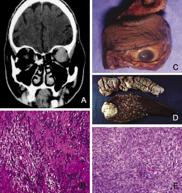
| Fig. 23 Schwannoma. The coronal CT scan shows a large well-delineated Schwannoma located in mid- and posterior orbit (A). Note the homogeneous appearance of the tumor with no bone infiltration. Histopathology from the same tumor reveals benign Schwannoma with Antoni type A tissue comprised of plump spindle cells arranged in interlacing bundles (B). Frame C shows an exenteration specimen with bone removal because of malignant Schwannoma. Metastatic malignant Schwannoma are depicted in pancreas and liver tissues from another case of craniofacial tumor (D). Frame E reveals the histopathology of malignant Schwannoma composed of pleomorphic spindle cells with hyperchromatic nuclei and numerous mitotic figures (arrows). |