|
|
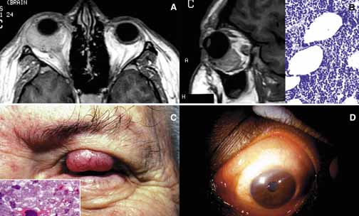
| Fig. 29 Lymphoma. Axial and sagittal T1-weighted images (A) of an orbital lymphoma that proved to be a maltoma histopathologically (B). Adipose tissue remains as oval, empty spaces within lymphoproliferative infiltration (B). Frame C reveals a large lymphoid lesion that occupies two-thirds of the left upper eyelid. Histopathologically, it was proven to be small cleaved cell lymphoma. The patient had systemic disease at the time of the development of his eyelid lesion. Frame D reveals the pinkish orange “salmon patch” lesion of a low-grade lymphoma involving the conjunctiva in a 360-degree fashion. The patient developed orbital lymphoma 3 years after diagnosis and treatment of the conjunctival lesion, but she was free of systemic disease. |