|
|
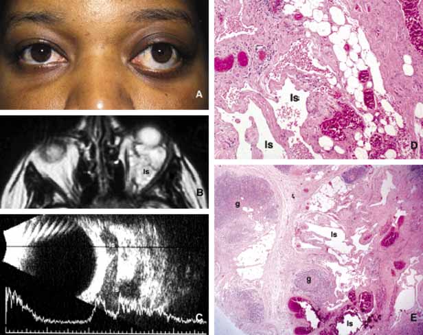
| Fig. 19 Lymphangioma. A 23-year-old woman with marked axial proptosis of the left eye secondary to lymphangioma (A). The tumor is depicted in the T1-weighted axial MRI showing a multiloculated tumor with marked enhancement occupying the entire orbit (B). Frame C shows multiple irregular lymphatic spaces (ls) that were changing in size and shape during the B-scan ultrasonography. The histopathology of the same case (D, E) reveals multiple lymphatic spaces (ls) lined by flat mesothelial-like cells surrounded with irregular fibroconnective tissue and lymphoid follicles with germinal centers (g). Note that some of the lymphatic spaces are filled totally or partially with blood. |