|
|
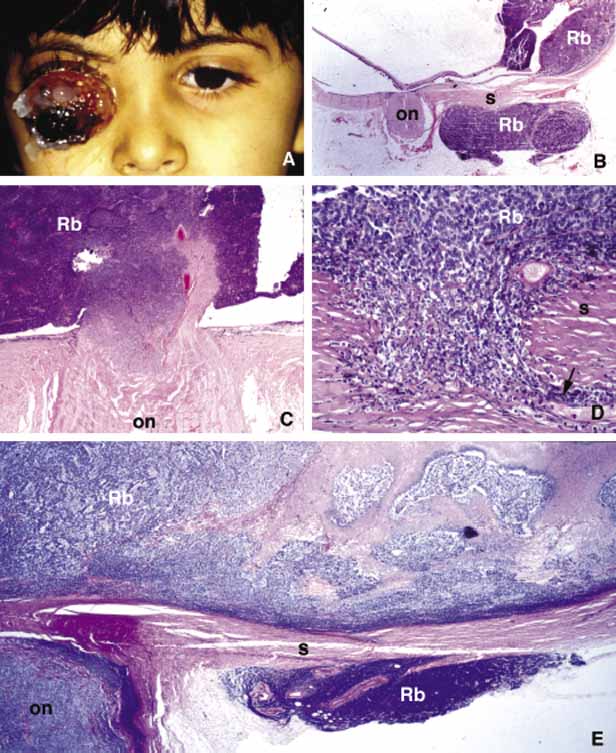
| Fig. 34 Orbital retinoblastoma. Frames A and B depict the anterior and posterior extraocular extension of retinoblastoma respectively. Frames C and D reveal the extension of retinoblastoma (Rb) into the optic nerve (on) and into scleral (s) emisserial channels. Frame E shows the low power microphotograph of the extension of retinoblastoma into the soft tissues of the orbit and the optic nerve (Rb, retinoblastoma; on, optic nerve; s, sclera). |