|
|
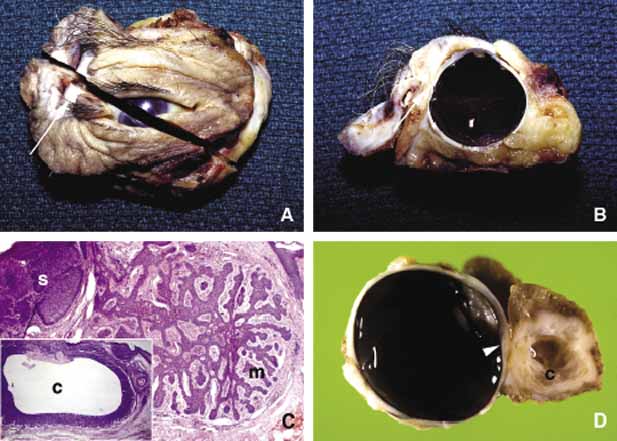
| Fig. 35 Basal cell carcinoma (BCC). Exenteration specimen (A, B) showing superior temporal basal cell carcinoma extending into the orbit and the globe (arrows). Frame C depicts different histopathologic types of basal cell proliferation in solid masses (s), in festoons (m), and as a cystic lesion (c). The basal cell proliferation forming ribbons and festoons turn into amorphoid type at the periphery. Frame D depicts a partial exenteration specimen showing a cystic BCC extending into the globe (arrowhead) and into the orbit posteriorly. |