|
|
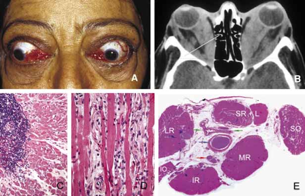
| Fig. 9 Graves disease. Clinical findings of Graves disease are depicted in frame A, including bilateral proptosis and lid lag with extraocular motility disturbance, chemosis with prolapse of lacrimal gland and congestion of conjunctival and episcleral blood vessels. Axial CT scan (B) reveals marked swelling of all recti muscles. The histopathologic appearance of the extraocular muscle from a patient with Graves disease reveals chronic inflammatory infiltrates, primarily composed of lymphocytes and plasma cells (C). The extraocular muscle volume is increased because of diffuse endomysial fibrosis, mucopolysacccharide deposition and chronic inflammatory cell infiltration (D). The orbital fat, meninges and the optic nerve (blue arrow), large blood vessels of the orbit, such as ophthalmic artery and its branches (green arrows) and the ciliary ganglion (red arrow) do not show any inflammation. The enlargement of the extraocular muscles are well depicted in frame E, which represents a transverse section, approximately at the level indicated by the yellow line in frame B. (LR: lateral rectus; SR + L: superior rectus and levator complex; SO: superior oblique; MR: medial rectus; IR: inferior rectus; IO: inferior oblique). (Frames C, D, and E are the courtesy of Ralph C. Eagle, MD of Philadelphia, PA) |