|
|
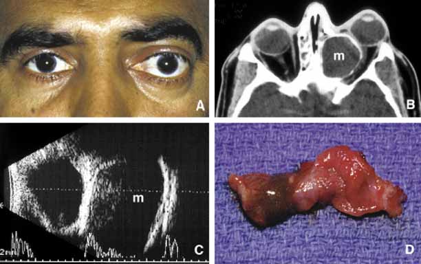
| Fig. 5 Mucocele. Moderate proptosis and slight lateral displacement of the left eye secondary to a large medially located mucocele (m) originating from the ethmoid sinus (A,B). Note the compression of the calcified wall of the lesion onto the globe and the optic nerve (B). The large cystic nature of the mucocele with low internal reflectivity and segmentally calcified wall is demonstrated by ultrasonography (C). Gross specimen of the stripped mucosal lining of the mucocele from the same case (D). |