|
|
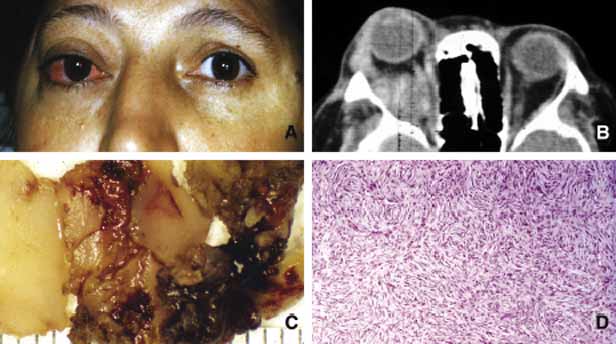
| Fig. 25 Fibrous histiocytoma. Proptosis and the congestion of the right eye secondary to an irregular space occupying lesion depicted on the axial CT scan (A, B). Note that the lesion is not well-delineated and compresses onto the optic nerve (B). Frame C depicts irregular fragments of the tissue obtained during the tumor debulking procedure. Note that some parts of the lesion are solid with homogenous appearance and other areas are more necrotic and hemorrhagic (C). Frame D illustrates the storiform appearance of spindle cells. In some cases these cells are accompanied by larger cells with bizarre or multiple nuclei. |