|
|
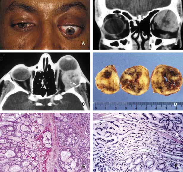
| Fig. 15 Adenoid cystic carcinoma. A large superior temporal mass causing marked proptosis of the eye and inferior displacement (A, B, C). The axial CT scan depicts the infiltration of the lateral wall with the tumor (arrow) (C). Serial sectioning of the tumor showing focal areas of hemorrhagic necrosis and cystic changes (D). Frames E and F depict the histopathological appearance of adenoid cystic carcinoma forming a “Swiss cheese” pattern with proliferation of atypical, irregular, glandular structures. Peripheral nerve sheath involvement (arrow) is seen in frame F. |