|
|
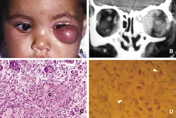
| Fig. 7 Orbital tuberculosis. A 2-year-old boy with left orbital tuberculosis (A). Both mother and child had systemic disease. A case of bilateral orbital and perinasal sinus tuberculosis is shown in the coronal CT of a 28-year-old man (B). Note the irregular involvement of the bony tissues of the orbits and the sinuses. C and D show a caseating granuloma (c) and AFB-positive tuberculous bacilli in necrotic inflammation (arrowheads) respectively. |