|
|
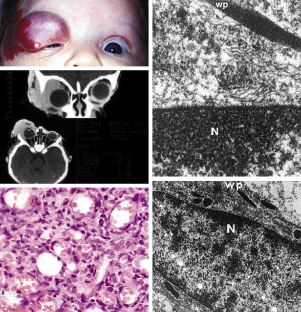
| Fig. 16 Capillary hemangioma. A large capillary hemangioma occupying the periorbital skin, upper eyelid and superior and inferior anterior orbit. The eye is dislocated inferiorly by the large tumor occupying the lateral and superior orbit. Note the lack of bone involvement on the CT scan despite the very large size of the tumor. The light microscopic appearance confirms the tumor as capillary hemangioma with proliferation of endothelial cells forming clusters and abnormal capillaries, some of which are developed well enough to contain red blood cells. The transmission electromicroscopy demonstrates in Weibel-Palade (wp) bodies to confirm the nature of the tumor cell as capillary endothelium [N, nucleus]. |