|
|
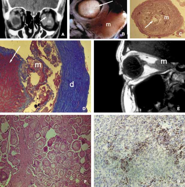
| Fig. 22 Meningioma. Coronal CT (A), sagittal T1-weighted MRI (E), gross specimen (B), and histopathologic photographs (C, D) show optic nerve meningiomas from different patients. In all cases, the optic nerve (white arrows) is pushed aside and/or compressed by the adjacent meningioma (m). In frames B, C, and D, the proliferation of the meningoendotheial cells (m) between dura (d) and the optic nerve (white arrows) are clearly seen. In psammomatous meningioma, meningoendothelial cells form numerous eddies surrounding partially calcified psammoma bodies (F). Immunohistochemically, meningioma is positive for EMA (G). |