|
|
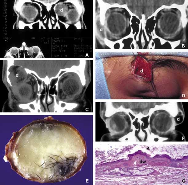
| Fig. 2 Dermoid. Different presentations of dermoid (d): superior medial, semi-solid mass pushing the globe down and out (A); a ruptured dermoid causing an inflammatory reaction within adjacent soft tissues (B); a large superior lateral dermoid eroding through the roof of the orbit to extend into the brain (C); extraorbital dermoid within the subcutaneous tissues of the eyebrow (D); gross appearance of the cystic dermoid containing whitish yellow cheesy keratin material intermixed with hair (E); dumbbell dermoid that is present on both sides of the frontozygomatic fissure (F); histopathology of dermoid wall (dw) containing skin appendages, the lumen of the dermoid is lined with stratified squamous epithelium producing keratin (K) (G). ([E] is the courtesy of Amin M. Nasr, MD of Beirut, Lebanon) |