|
|
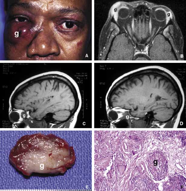
| Fig. 11 Sarcoidosis. The appearance of a large sarcoidosis granuloma involving the anterior orbit and periorbital skin (g). The central dome-like lesion is caused by secondary to a Staphylococcus infection. In addition, plaquoid skin lesions of sarcoidosis are seen above the left eyebrow, on the left upper eyelid, and on the periorbital skin (A). The T2-weighted axial MRI with contrast reveals bilateral enlargement of lacrimal glands (g) in another case B. Frames C and D show T1-weighted sagittal MRIs with localized granulomatous masses of sarcoidosis within the posterior orbit involving the apex and extending into the cavernous sinus (C, D). A well-delineated but not encapsulated mass of a sarcoid granuloma (E). The histopathology of sarcoidosis consists of multiple granulomas composed of histiocytic cells, chronic inflammatory cells, and multinucleated giant cells (g) (F). |