|
|
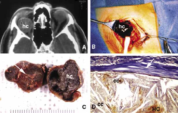
| Fig. 4 Hematic cyst. Axial CT scan (A) showing a large superiorly located, well-circumscribed hematic cyst (hc) presenting as an homogeneous low-density image. Intraoperative photograph of the same case shows dark brown hematic cyst. (B). C and D show the gross and histopathologic appearance of the hematic cyst respectively. It is surrounded by a fibrous pseudocapsule (arrows) containing a mixture of cholesterol crystals, (cc), hematoidin crystals (HC), and other proteineceous debris. |