|
|
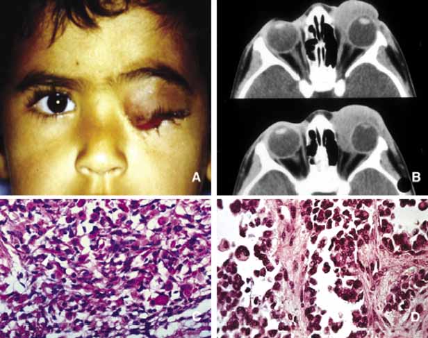
| Fig. 24 Orbital rhabdomyosarcoma. Frames A and B depict anterior medial orbital rhabdomyosarcoma that was histopathologically proven to be of embryonal type composed of neoplastic skeletal muscle cells with round to oval hyperchromatic nuclei and abundant pink cytoplasms. Elongated and tadpole-shaped cells may be seen. The identification of intracytoplasmic cross striations confirms the origin of the tumor as skeletal muscle (C). Frame D depicts the histopathology of the alveolar rhabdomyosarcoma with the distinctive architecture in which the cells are interspersed as a branching network of fibroconnective tissue. The alveolar type presents with larger and more pleomorphic appearing tumor cells. (Frame C is the courtesy of Doris Hadjistilianou, MD of Siena, Italy) |