|
|
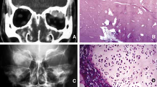
| Fig. 27 Bone tumors. Coronal CT scan shows an oval-shaped osteoma originating from markedly thickened bone of the orbital roof (A). Note that although the tumor is benign it presents with irregular margins that blend into mature cavernous bone; this is well-depicted histologically in frame B. Anterior-posterior view of a plain skull film shows a large irregular density of a chondrosarcoma originating from the lateral aspect of the orbit (C). The light microscopic appearance of the tumor reveals numerous clusters of moderately pleomorphic hyaline cartilage cells forming lobules (D). |