|
|
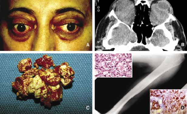
| Fig. 26 Orbital xanthogranulomatosis (xg). Bilateral proptosis, frozen orbits secondary to massive infiltration with partially necrotic xanthogranulomatous lesions (A, B). Note the yellow xanthoma plaques on the upper eyelids and within the medial canthal areas (A). Frame C depicts multiple fragments of partially hemorrhagic xanthogranulomatous tissue at the time of surgery. Same patient also had systemic changes of Erdheim-Chester disease with long bone involvement, abnormal EKG, and a retroperitoneal mass (D). Insets depict large foamy histiocytes stained with H E (upper inset) and Oil red-0 (lower inset) stains. Note the extensive fat staining (red) of the lesion. |