|
|
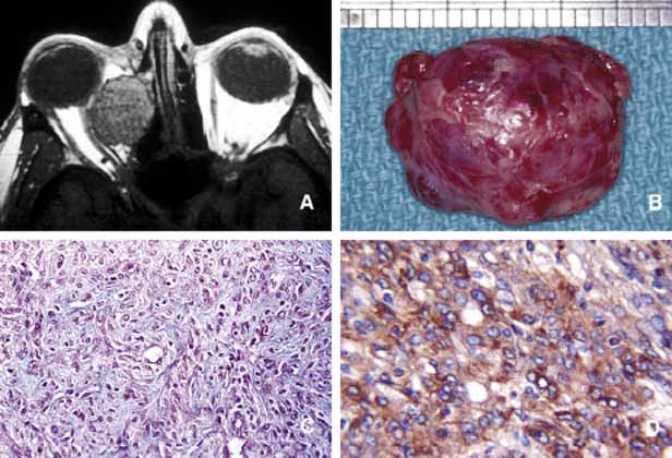
| Fig. 17 Hemangiopericytoma. Axial T1-weighted MRI showing a well-encapsulated large mass located in the medial orbit with compression onto the globe and the optic nerve (A). The signal intensity varies within the mass because of its vascular nature. Frame B depicts the well-encapsulated hemangiopericytoma at the time of its removal. Microscopic appearance reveals a mixture of haphazardly arranged spindle-shaped tumor cells with round and oval nuclei and scanty cytoplasms, mixed with a network of sinusoidal spaces and/or abnormally developed blood vessels. Moderate degree of pleomorphism and occasional abnormal mitotic figures are seen in this section (C). Although these malignant histopathology indicators have been reported in tumors that later metastasize; as a rule the biologic behavior of hemangiopericytoma cannot be dependably assessed based on its histopathology. Frame D shows the immunohistochemical staining with vimentin. |