|
|
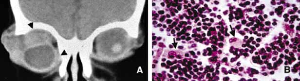
| Fig. 33 Neuroblastoma. The coronal CT scan showing bilateral metastatic tumors with a much larger mass in the right orbit (A). Note the indentation and inferior dislocation of the right globe secondary to superiorly located tumor. Metastatic neuroblastoma is well-known for its haphazard infiltration into the bone; however, in this case, although some irregularities can be seen in the superior and medial orbital walls (arrowheads), obvious bony involvement was not present. Histopathologically neuroblastoma consists of sheets of small tumor cells with round and oval hyperchromatic nuclei (B). In between there are a few differentiating neuroblasts with vesicular nuclei and prominent neucleoli (arrows). |