|
|
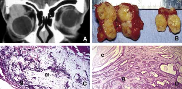
| Fig. 14 Benign mixed tumor (BMT). Coronal CT (A) depicts a well-delineated mass in the superior lateral orbit causing inferior medial dislocation of the globe. Frame B shows multilobulated nature of pleomorphic ademona (benign mixed tumor). The tumor nodules are bisected to show the encapsulation and the focal nature of the tumor masses; the nodule on the right was discovered inferior to the main lesion as a satellite mass (B). Histopathologically pleomorphic adenoma is composed of a mixture of glandular (g) and myxoid (m) tissues (C, D). Some of the glandular formations may develop squamous metaplasia and distend to form cystic space (c) secondary to keratin accumulation. (D) |