|
|
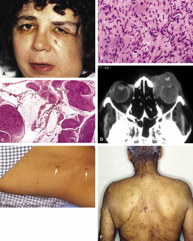
| Fig. 20 Neurofibromatosis. The deformed face of a patient with neurofibromatosis of the orbit, eyelids, and facial skin. (A) The histopathology of plexiform neurofibroma is composed of bundles of neoplastic nerve fiber intermixed with collagen in a myxomatous matrix and fibroadiopose tissue (B). Frame C depicts a high-power magnification with neurofibroma composed of a mixture of formed cells with wavy nuclei and neurons intermixed within collagenous tissue (C). An axial CT scan of a patient with an anteriorly located plexiform neurofibroma (nf) (D). Frames E and F show skin changes of neurofibromatosis type I including pigmented neurofibromas, café au lait spots, and pigmented freckles in the axilla (E). |