Chapter 113A
Fluorescence Angiography of Choroidal and Retinal Tumors
JAMES J. AUGSBURGER
Main Menu   Table Of Contents

Search

CHOROIDAL TUMORS
RETINAL TUMORS
SUMMARY
REFERENCES

Fluorescence angiography has been used extensively in the evaluation of tumors of the choroid and retina during the past four decades. Several excellent reviews on the subject of fluorescein and indocyanine green (ICG) angiography of intraocular tumors have been published,1, 2, 3, 4, 5, 6, 7 and interested readers are referred to these sources for additional information on this subject. The role of fluorescence angiography in a patient with an intraocular tumor is largely documentary in nature, but it can be of differential diagnostic value in certain cases. The essential clinical importance of fluorescence angiography in the evaluation of most choroidal and retinal tumors appears to be its ability to reveal patterns inconsistent with the presumptive diagnosis rather than to elucidate pathognomonic features.8 The role and importance of fluorescence angiography in various tumors of the choroid and retina are summarized in the remainder of this chapter.
Back to Top
CHOROIDAL TUMORS
Both fluorescein and ICG angiography are used currently to evaluate many choroidal tumors. Because ICG angiography tends to image choroidal blood vessels substantially better than does fluorescein angiography, it appears to be the preferred method for evaluating most choroidal tumors. However, because fluorescein angiography has been used more extensively, both the fluorescein and ICG angiographic features of the following selected choroidal tumors are described and illustrated.

CHOROIDAL NEVUS

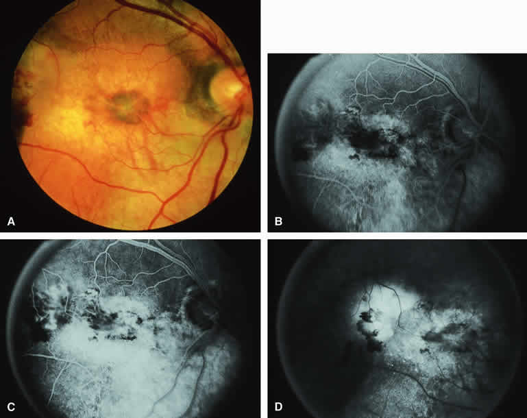
The choroidal nevus is a benign uveal melanocytic tumor of limited growth potential. The typical lesion (Figs. 1, 2, 3, and 4) appears as an ill-defined, gray-brown choroidal mass that is usually less than 5 mm in maximal basal diameter and less than 1 mm in thickness. The basal margins of typical lesions often blend into the normal surrounding choroid in a feathered or striate pattern. Drusen and retinal pigment epithelial (RPE) pigment clumps commonly develop on the surface of these tumors.

Fig. 1. Typical melanotic choroidal nevus. A. Ill-defined gray-brown macular choroidal lesion with bland surface features. B-D. Fluorescein angiogram of lesion. B. Laminar venous phase frame showing intact retinal vasculature overlying nevus but virtually no hypofluorescence corresponding to choroidal lesion. C. Full venous phase frame showing granular hyperfluorescence corresponding to minor retinal pigment epithelial alterations and drusen on surface of tumor. D. Late-phase frame showing continued hypofluorescence of choroidal lesion with mild staining of surface drusen.

Fig. 2. Typical melanotic choroidal nevus. A. Small gray choroidal nevus one disc diameter above fovea. The pale lesion at upper left margin of photograph is a choroidal melanoma that was the main focus of the following angiogram. B-D. Indocyanine green (ICG) angiogram of lesion. B. Early-phase frame showing intense hypofluorescence of nevus. C. Intermediate-phase frame showing sustained hypofluorescence of nevus. D. Late-phase frame showing sustained hypofluorescence of nevus.

Fig. 3. Amelanotic choroidal nevus. A. Ill-defined pale yellow choroidal nevus superior to optic disc. B-D. Indocyanine green (ICG) angiogram of lesion. B. Early-phase frame showing mild hypofluorescence of nevus and visibility of large-caliber choroidal arteries passing through lesion. C. Intermediate-phase frame showing features similar to those of the previous frame. D. Later phase frame showing ill-defined mild hyperfluorescence of lesion.

Fig. 4. Melanotic choroidal nevus with drusen and retinal pigment epithelial (RPE) alterations. A. Gray-brown choroidal nevus with numerous white drusen and dark gray pigment clumps on its surface. B-D. Fluorescein angiogram of lesion. B. Venous phase frame showing retinal arteries and veins but no convincing sign of the underlying choroidal nevus. C. Later venous phase frame showing mild granular hyperfluorescence and hypofluorescence centrally corresponding with drusen and RPE pigment clumps overlying choroidal nevus. D. Late venous phase frame showing increased multifocal hyperfluorescence corresponding to drusen and persistent foci of hypofluorescence corresponding to RPE alternations.

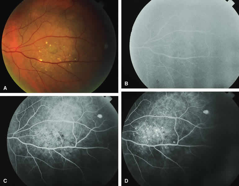
Fluorescein and ICG angiographic features of choroidal nevi have been described by numerous authors.1, 2, 3, 4, 5, 6, 7, 9, 10, 11, 12 Neither fluorescein nor ICG angiography appears to be particularly helpful for evaluation of a typical choroidal nevus. Ophthalmoscopy is usually sufficient to allow one to decide with reasonable certainty whether a small melanotic choroidal tumor is a typical choroidal nevus or an equivocal lesion that may be either a large nevus or small choroidal melanoma. No well-designed clinical study has ever demonstrated significant independent differential diagnostic value of fluorescence angiography for clarifying this differential diagnosis.8

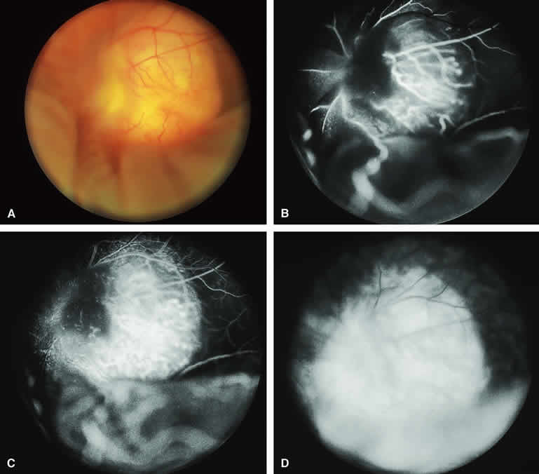
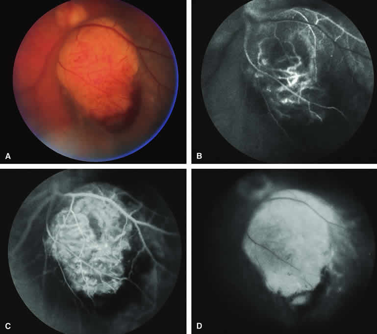
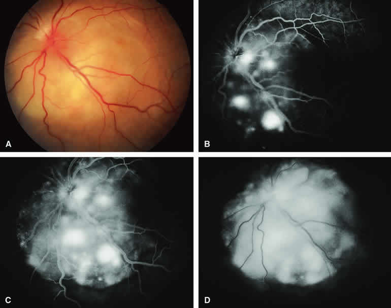
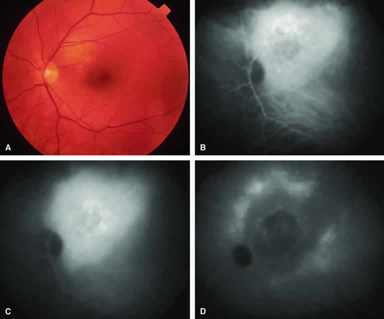
Typical Melanotic Choroidal Nevus

Fluorescein angiography of a typical choroidal nevus with bland surface features (see Fig. 1) shows the entire lesion to be hypofluorescent relative to the adjacent uninvolved choroid throughout the study. No large-caliber choroidal blood vessels are usually identifiable within the lesion. The retinal vasculature overlying the lesion appears well defined and normal on fluorescein angiography.

ICG angiography of a typical melanotic choroidal nevus (see Fig. 2) shows better definition of the basal area of the lesion than does fluorescein angiography. The entire lesion appears completely and uniformly dark throughout the ICG angiogram. Only the larger retinal blood vessels overlying the nevus are usually demonstrated on ICG angiography.

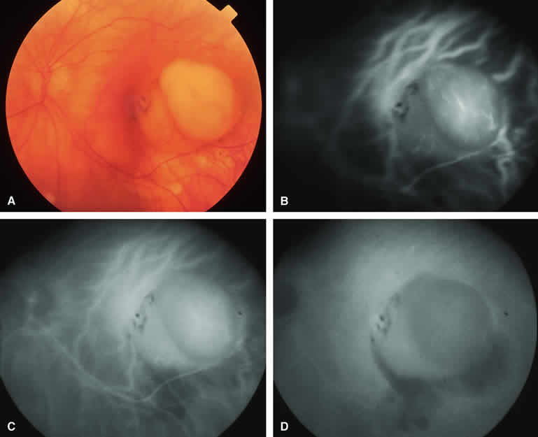
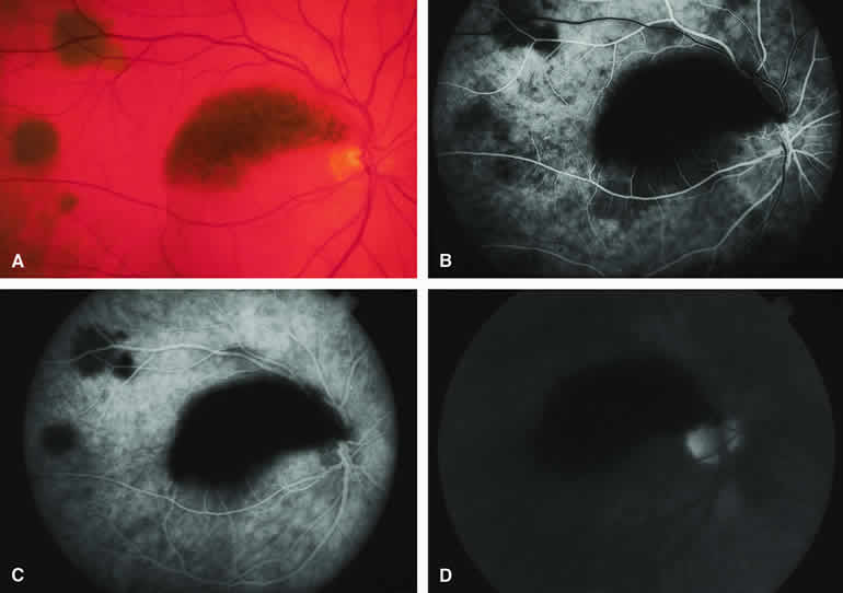
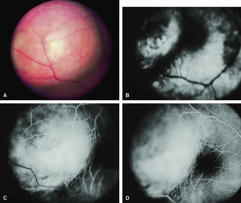
Amelanotic Choroidal Nevus

Approximately 10% to 15% of choroidal nevi are largely or completely amelanotic clinically. Fluorescein and ICG angiography of an amelanotic choroidal nevus (see Fig. 3) tend to show less prominent hypofluorescence of the lesion than they do with darkly melanotic nevi. Because of the lack of intracellular melanin pigment within the nevus cells, some large-caliber choroidal blood vessels running through the nevus may be visible in the region of the mass (see Fig. 3B and C). These choroidal blood vessels are better defined by ICG angiography than by fluorescein angiography. Amelanotic choroidal nevi often appear mildly hyperfluorescent in late-phase frames (see Fig. 3D).

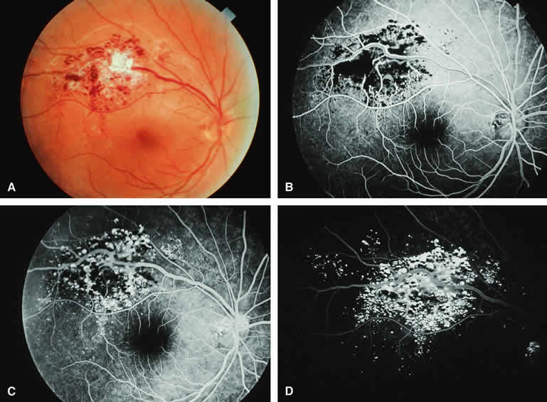
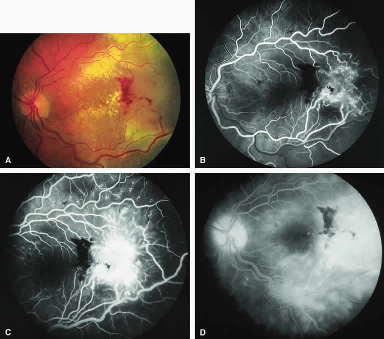
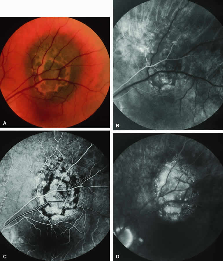
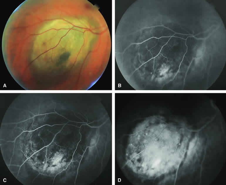
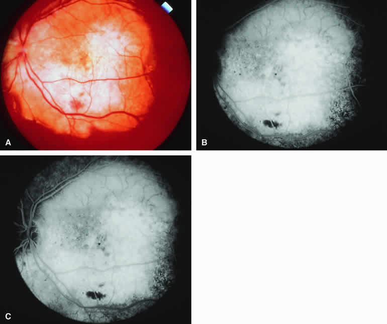
Choroidal Nevus with Drusen and Clumps of RPE Hyperplasia

If a choroidal nevus has drusen and RPE alterations on its surface (see Fig. 4A), fluorescein angiography (Fig. 4B, C, and D) tends to show patchy or stippled window defect hyperfluorescence corresponding to foci of RPE depigmentation, fluorescence blockage by clumps of RPE hyperplasia on the surface of the lesion, and late staining of at least some of the drusen. These features are not usually as evident on ICG angiography as they are on fluorescein angiography.

Choroidal Nevus Versus Melanoma

Small melanocytic choroidal tumors larger than 5 mm in diameter and thicker than 1 mm but without clearly invasive clinical features (e.g., nodular eruption through Bruch's membrane, retinal invasion) may be either benign nevi or small choroidal melanomas. Several features of these lesions have been identified as prognostic of the likelihood of subsequent lesion enlargement,13 a surrogate indicator of the malignant potential of the tumor. Features suggestive of low growth potential and probable benign histology include thickness less than or equal to 1.5 mm, uniform gray-brown coloration of the lesion, and drusen and RPE clumping on the surface of the lesion. In contrast, features suggestive of higher growth potential and probable malignant histology include thickness greater than 1.5 mm, nonuniform coloration of the lesion, prominent clumps of lipofuscin pigment on the surface of the lesion, and serous subretinal fluid overlying and surrounding the lesion. None of these features is a reliable indicator of the underlying histologic nature of the tumor; however, the greater the number of unfavorable features, the greater the likelihood of lesion enlargement if it is followed without treatment after initial detection.

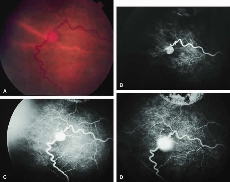
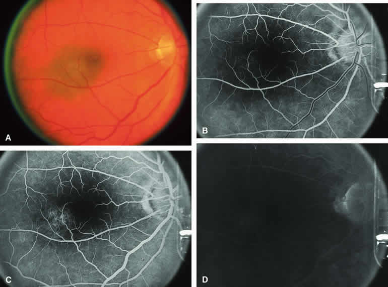
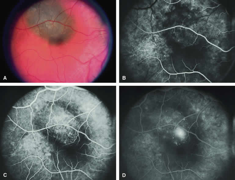
Choroidal Nevus Versus Melanoma with Bland Surface Features

Several authors have evaluated fluorescein and ICG angiography as differential diagnostic tools for differentiating between large benign choroidal nevi and small malignant choroidal melanomas.9, 10 An example of such a lesion evaluated by fluorescein angiography is presented as Figure 5. Some common assertions based on these studies include the following. If the lesion is a nevus, fluorescein angiography is likely to show hypofluorescence of the lesion throughout the study, absence of large-caliber intralesional blood vessels, and lack of late smudgy hyperfluorescence resulting from leakage from these vessels. In contrast, if the lesion is a melanoma, fluorescein angiography is likely to show at least some intralesional tumor blood vessels, some patchy or smudgy fluorescein leakage from those blood vessels, and at least patchy or smudgy late fluorescence of the tumor. ICG angiography is more likely to reveal intralesional blood vessels within bland appearing small melanocytic choroidal lesions (especially darkly melanotic lesions) than is fluorescein angiography, and lesions with prominent intralesional blood vessels are generally regarded as more likely to be melanomas. Unfortunately, there have been no confirmatory angiographic-histopathologic correlation studies of small melanocytic choroidal tumors in the nevus versus melanoma category to confirm or refute these assertions. At best, this author regards the fluorescence angiographic features of bland appearing small melanocytic choroidal tumors as suggestive but not confirmatory of the pathologic nature of the tumor.

Fig. 5. Choroidal nevus versus melanoma. A. Partially melanotic and partially amelanotic choroidal tumor with prominent clumps of retinal pigment epithelial (RPE) pigment and drusen on surface. B-D. Fluorescein angiogram of lesion. B. Laminar venous phase frame showing poorly defined hypofluorescence of choroidal lesion and area of window defect hyperfluorescence corresponding to amelanotic portion of lesion. C. Full venous phase frame showing persistent mild hypofluorescence corresponding to melanotic portion of lesion, increased hypofluorescence corresponding to RPE thinning and drusen overlying lesion, and foci of intense hypofluorescence corresponding to RPE clumps on lesion. D. Late-phase frame showing persistence of features shown in the prior frames plus more numerous hyperfluorescent foci corresponding with additional drusen.

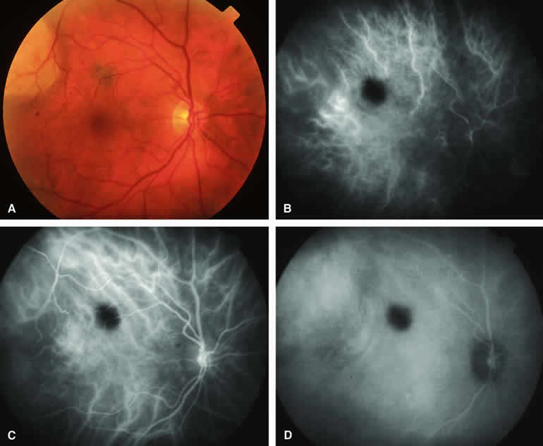
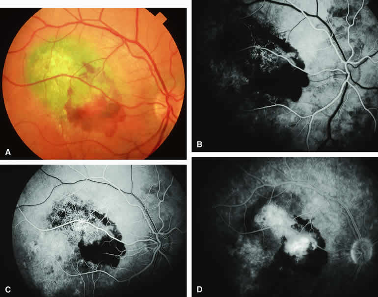
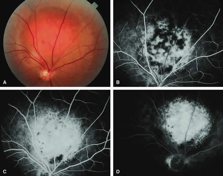
Choroidal Nevus Versus Melanoma with Prominent Lipofuscin Pigment Clumps

If one obtains a fluorescein angiogram on a small melanotic choroidal lesion (nevus versus melanoma) that has prominent clumps of lipofuscin pigment on its surface (Fig. 6), the pigment clumps appear intensely hypofluorescent throughout the study. This appearance is attributable to the complete blocking of choroidal fluorescence by the lipofuscin. ICG angiography does not show lipofuscin pigment clumps on the surface of the tumor as well as fluorescein angiography does.

Fig. 6. Melanotic choroidal nevus versus melanoma with prominent clumps of overlying orange pigment. A. Darkly melanotic choroidal lesion with prominent clumps of overlying orange pigment (lipofuscin). B-D. Fluorescein angiogram of lesion. B. Arterial phase frame showing irregular hypofluorescence and hyperfluorescence corresponding to lesion. C. Laminar venous phase frame showing generalized window defect hyperfluorescence of mass plus intense choroidal fluorescence blockage by orange pigment. D. Late-phase frame showing multiple pinpoint foci of hyperfluorescence at retinal pigment epithelial (RPE) level, leakage of fluorescein into the overlying and surrounding subretinal fluid, and partial obscuration of the blockage of choroidal hypofluorescence corresponding to the orange pigment on the surface of the lesion.

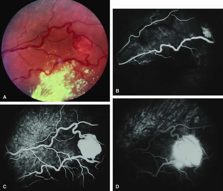
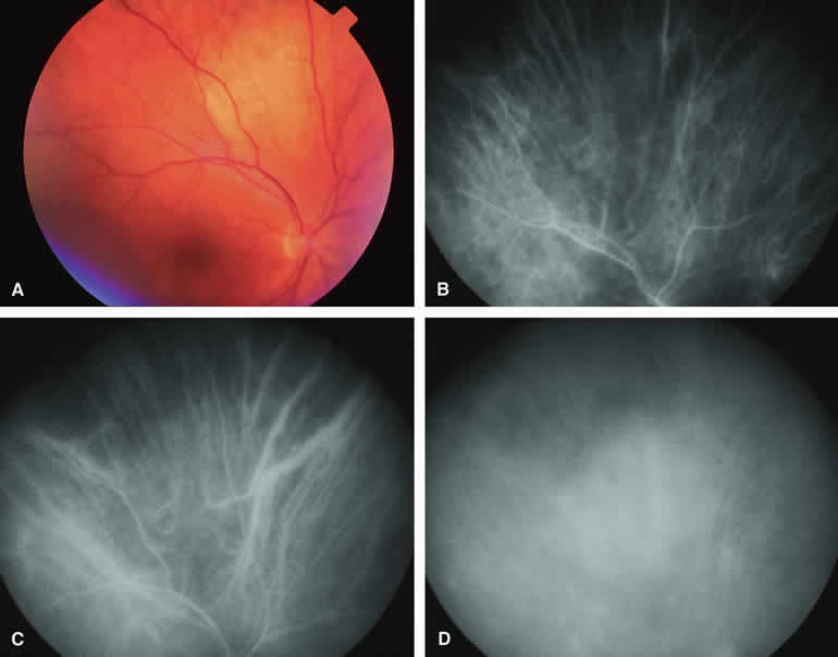
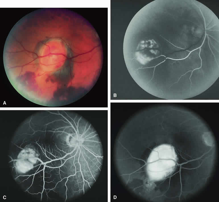
Choroidal Nevus Versus Melanoma with Overlying Serous Subretinal Fluid

A blister of serous subretinal fluid sometimes develops over and around a presumed choroidal nevus (Fig. 7A), especially if the lesion is located in the macula.11 If one performs a fluorescein angiogram on a small melanocytic choroidal lesion (nevus versus melanoma) that has shallow overlying serous subretinal fluid (see Fig. 7B, C, and D), one or more hyperfluorescent leak sites may show up slowly at the RPE level as the study progresses. In some cases, fluorescein will clearly leak from those foci into the overlying serous subretinal fluid. ICG angiography does not show hyperfluorescent leak sites at the RPE level as well as fluorescein angiography does.

Fig. 7. Melanotic choroidal nevus versus melanoma with overlying serous subretinal fluid. A. Bland gray-brown choroidal lesion involving upper portion of macula with shallow overlying and surrounding blister of serous subretinal fluid. B-D. Fluorescein angiogram of lesion. B. Arterial phase frame showing patchy choroidal hypofluorescence corresponding to choroidal lesion. C. Full venous phase frame showing faint zone of choroidal hypofluorescence corresponding to marginal zone of lesion, mild hyperfluorescence corresponding to the central portion of the lesion, and discrete pinpoint foci of hyperfluorescence at the retinal pigment epithelial (RPE) level on the lesion. D. Later phase frame showing smudge hyperfluorescent focus of fluorescein leakage into subretinal fluid.

Choroidal Nevus Versus Melanoma with Choroidal Neovascular Membrane

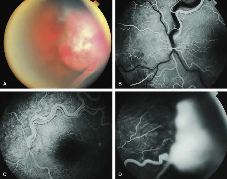
A choroidal neovascular membrane occasionally develops from the surface of a small melanocytic choroidal tumor (nevus versus melanoma).12 This vascular structure can usually be anticipated because of the presence of ophthalmoscopically evident hemorrhagic or exudative subretinal fluid overlying a portion of the tumor (Fig. 8A). Fluorescein angiography in such cases (see Fig. 8B to D) generally reveals the neovascular membrane as a relatively well-defined vascular network that fluoresces brightly in the early frames of the study and leaks progressively by the late frames. If the subretinal fluid is grossly hemorrhagic, ICG angiographymay show the choroidal neovascular network better than does fluorescein angiography.

Fig. 8. Melanotic choroidal nevus versus melanoma with overlying choroidal neovascular membrane. A. Same lesion shown in Figure 7 following spontaneous accumulation of subretinal blood and hard exudates associated with development of choroidal neovascular membrane overlying lesion. B-D. Fluorescein angiogram of lesion. B. Arterial phase frame showing abnormal vascular network deep to retina, partially obscured by blockage corresponding to subretinal blood. C. Laminar venous phase frame showing greater extent of abnormal vascular network overlying lesion and zone of window defect hyperfluorescence extending inferiorly through the macula. D. Late-phase frame showing smudgy hyperfluorescence from abnormal subretinal blood vessels, persistent blockage of choroidal fluorescence by the subretinal blood, and persistence of later hyperfluorescence due to retinal pigment epithelial (RPE) thinning extending inferiorly through the macula.

Fluorescence Angiographic Detection of Lesion Enlargement

Small melanocytic choroidal lesions in the nevus versus melanoma category are frequently monitored periodically following initial documentation for enlargement or other indicators of possible malignant behavior. Fluorescein angiography sometimes defines the basal area of these lesions better than does fundus photography, but ICG angiography is clearly the preferred technique for this purpose. If one elects to follow a small melanocytic choroidal tumor without treatment, comparative fluorescence angiography can be used to identify slight lesion enlargement that is not clearly evident by ophthalmoscopy or comparison of fundus photographs. Because benign choroidal nevi can and do enlarge, however, detection of subtle lesion enlargement by fluorescence angiography probably has limited differential diagnostic value in these lesions.

CHOROIDAL MALIGNANT MELANOMA

Choroidal malignant melanoma is the most common primary malignant intraocular neoplasm of adults. It has substantial local growth potential in addition to its well-known propensity to metastasize and, thereby, prove fatal to the host. Fluorescence angiography can provide evidence in support of the diagnosis of choroidal melanoma but is rarely if ever sufficient by itself to establish the diagnosis. Several ophthalmoscopically detectable features of clinically diagnosed melanocytic choroidal tumors are strongly indicative of their malignant potential. These include tumor diameter substantially greater than 7 mm, tumor thickness substantially greater than 3 mm, prominent intralesional large-caliber blood vessels, nodular apical eruption of the tumor through Bruch's membrane, invasion of the overlying retina by the tumor, and a prominent nonrhegmatogenous retinal detachment with shifting subretinal fluid.

Several distinct fluorescein and ICG angiographic patterns have been associated with choroidal melanomas.1, 2, 3, 4, 5, 6, 7, 14, 15, 16, 17, 18, 19, 20, 21 These distinct patterns depend on the degree of pigmentation of the tumor cells, the thickness of the tumor, the presence or absence of eruption of tumor through the overlying Bruch's membrane, and the presence or absence of retinal invasion by the tumor.

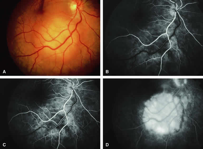
Melanotic Choroidal Melanoma Without Invasive Features

The typical choroidal melanoma that has not broken through Bruch's membrane generally appears as a gray-brown to dark brown, dome-shaped, solid choroidal mass (Fig. 9A). The overlying retina commonly appears normal except that it is draped over the choroidal tumor. Fluorescein angiography (see Fig. 9B, C, and D) typically shows the tumor to be relatively hypofluorescent during the early frames of the study. If prominent clumps of orange lipofuscin pigment are present on the surface of the tumor, they will appear intensely hypofluorescent throughout the study because of blockage of the underlying choroidal and tumor vascular fluorescence. During the arterial phase frames of the study, several ill-defined large-caliber deep intralesional blood vessels may be identifiable against the generally hypofluorescent background. As the study continues, these large intralesional blood vessels leak progressively so that the surface of the lesion tends to appear hyperfluorescent by the late frames. Fluorescein may also accumulate in the overlying retina and retinal pigment epithelium as tiny discrete pinpoint foci of intense hyperfluorescence. If serous retinal detachment is present overlying and around the tumor, the fluorescein will leak through the retinal pigment epithelium and accumulate in the subretinal fluid by the late frames. The retinal vascular pattern of the overlying retina commonly appears unremarkable in these cases.

Fig. 9. Typical melanotic choroidal melanoma without invasive features. A. Nodular gray-brown nodular choroidal tumor with linear clumps of orange lipofuscin pigment and central whitish discoloration of overlying retinal pigment epithelium. B-D. Fluorescein angiogram of lesion. B. Venous phase frame showing mild hypofluorescence of most of tumor but with ill-defined hyperfluorescent focus near inferior margin. C. Later venous phase frame showing persistent generalized hypofluorescence of mass, increased smudgy and punctate hyperfluorescence near the inferior and temporal margins of lesion, and choroidal fluorescence blockage by lipofuscin pigment overlying mass. D. Late-phase frame showing diffuse hyperfluorescence of subretinal fluid and outer retina overlying mass, persistent hypofluorescence corresponding to the margin of the lesion, persistent choroidal fluorescence blockage corresponding to the lipofuscin pigment clumps, and several pinpoint dots of intense hyperfluorescence overlying the mass.

If ICG angiography is performed on a similar tumor (Fig. 10), the tumor mass generally appears more intensely hypofluorescent throughout the study and its intrinsic blood vessels appear more clearly visible than on fluorescein angiography. ICG slowly accumulates within the extracellular space of the tumor so that the tumor usually appears mildly hyperfluorescent, at least in part, in the late frames.

Fig. 10. Typical melanotic choroidal melanoma without invasive features. A. Ill-defined bilobed melanotic choroidal mass with prominent orange lipofuscin pigment clumps on its surface. B-D. Indocyanine green (ICG) angiogram of lesion. B. Early-phase frame showing generalized hypofluorescence of mass but with large-caliber choroidal blood vessels passing through it. C. Later phase frame showing persistent hypofluorescence and better definition of choroidal lesion. Some choroidal blood vessels are still visible through the mass. The lipofuscin pigment clumps produce limited choroidal fluorescence blockage. D. Late-phase frame showing persistent hypofluorescence blockage of most of mass but some smudgy hyperfluorescence of marginal aspects of lesion.

Amelanotic Choroidal Melanoma Without Invasive Features

If the choroidal melanoma being evaluated is relatively amelanotic (Figs. 11A and 12A), its large-caliber intralesional blood vessels will tend to show up more distinctly on both fluorescein angiography (see Fig. 11B, C, and D) and ICG angiography (see Fig. 12B, C, and D) than they would in a darkly melanotic choroidal melanoma. Despite its amelanotic color, the cellular component of the mass tends to be at least mildly hypofluorescent relative to the adjacent uninvolved choroid during the early frames of the study. As with melanotic melanomas, the mass usually appears at least mildly hyperfluorescent in the late-phase frames (see Figs. 11D and 12D).

Fig. 11. Amelanotic choroidal melanoma without invasive features. A. Amelanotic, golden-yellow choroidal tumor with prominent visibility of intralesional blood vessels. B-D. Fluorescein angiogram of lesion. B. Choroidal filling phase frame showing generalized hypofluorescence of lesion but with well-defined intralesional large-caliber blood vessels not connected to retinal vasculature. C. Retinal arterial phase frame showing even more prominent intralesional large blood vessels against persistently hypofluorescent tumor. D. Late-phase frame showing diffuse hyperfluorescence of surface of lesion resulting from exuberant leakage of fluorescein into subretinal space and lesion.

Fig. 12. Amelanotic choroidal melanoma without invasive features. A. Pale yellow macular choroidal tumor with tiny clumps of disrupted retinal pigment epithelial (RPE) pigment on its surface. B-D. Indocyanine green (ICG) angiogram of lesion. B. Early-phase frame showing prominent fluorescent large-caliber intralesional blood vessels against the background of a generally hypofluorescent choroidal mass. C. Later phase frame showing persistent hypofluorescence of the mass except for the prominent intralesional blood vessels. D. Late-phase frame showing increase in fluorescence of mass compared with surrounding normal choroid and marginal hyperfluorescence resulting from ICG accumulation in the overlying subretinal fluid.

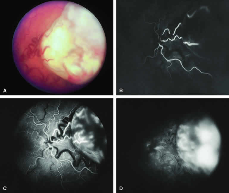
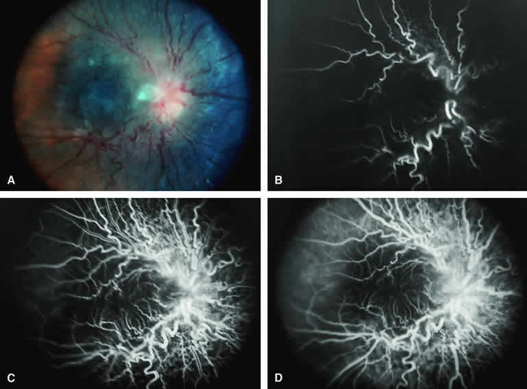
Choroidal Melanoma With Nodular Eruption Through Bruch's Membrane

If a choroidal melanoma has erupted through Bruch's membrane (Figs. 13A and 14A), it forms an apical nodule that is generally hypomelanotic and contains many large-caliber blood vessels. Fluorescein angiography of these tumors (see Fig. 13B, C, and D) typically shows hypofluorescence of the base of the lesion during the early frames, relatively rapid filling of the prominent blood vessels in the apical nodule during the venous and recirculation frames, and intense late staining of the apical nodule resulting from progressive fluorescein leakage by the late-phase frames. Similarly, ICG angiography of these tumors (see Fig. 14B, C, and D) also shows relative early hypofluorescence of the tumor base, early filling of prominent intralesional blood vessels within the apical nodule, intense staining of the apical nodule by the recirculation phase frames, and persistent late hyperfluorescence of the mass.

Fig. 13. Choroidal melanoma with nodular eruption through Bruch's membrane. A. Mushroom-shaped choroidal melanoma inferonasal to optic disc, showing dark basal region and lighter apical eruption through Bruch's membrane. Note prominence of intralesional blood vessels within apical portion of lesion. B-D. Fluorescein angiogram of lesion. B. Arterial phase frame showing fluorescence of large-caliber blood vessels within hypofluorescent apical nodule of tumor. C. Venous phase frame showing increased prominence of intralesional blood vessels, as well as alterations of retinal capillary bed where retina is thinned over apex of tumor. D. Late-phase frame showing intense hyperfluorescence of apical nodule of tumor.

Fig. 14. Choroidal melanoma with nodular eruption through Bruch's membrane. A. Amelanotic small macular choroidal tumor with amelanotic apical nodular eruption through Bruch's membrane and clumps of black retinal pigment epithelial (RPE) pigment along nasal margin of lesion.B-D. Indocyanine green (ICG) angiogram of lesion. B. Early-phase frame showing several prominent fluorescent intralesional blood vessels within apical nodule, generalized mild hypofluorescence of lesion, and choroidal fluorescence blockage by the RPE pigment clumps on the subfoveal portion of the lesion's base. C. Intermediate-phase frame showing increased fluorescence of tumor and reduced visibility of intralesional blood vessels. D. Late-phase frame showing greater hyperfluorescence of tumor base than of apical nodule and area of hypofluorescence along inferior margin of mass.

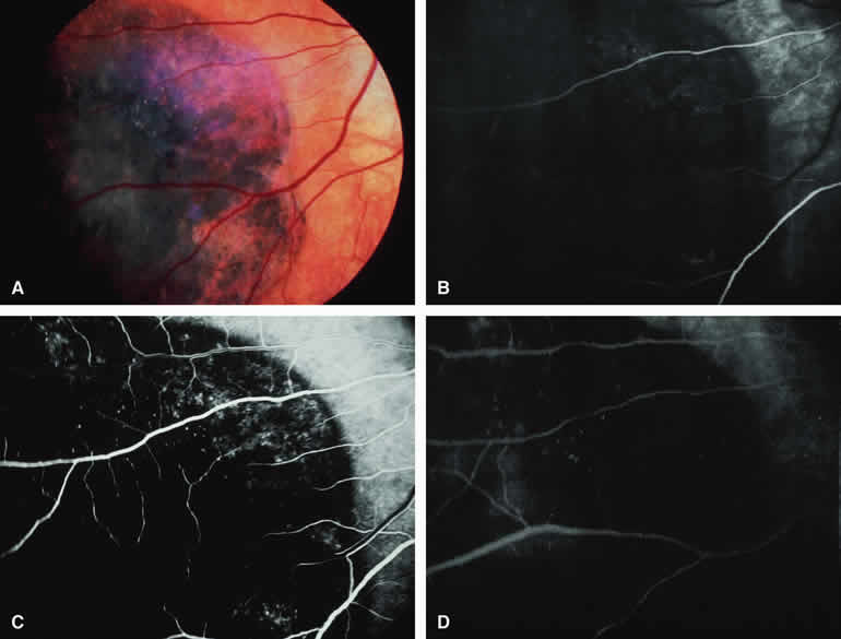
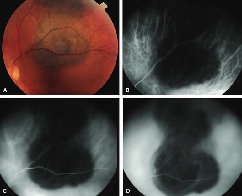
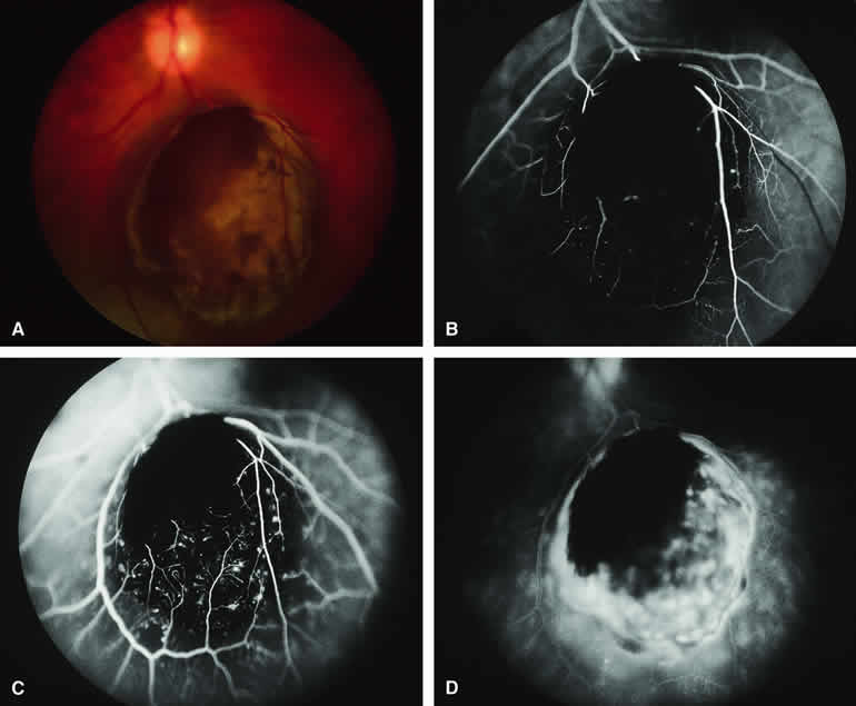
Choroidal Melanoma with Retinal Invasion

Occasional choroidal melanomas develop darkly melanotic patches of retinal invasion on their surface. These patches appear as homogeneous dark brown velvety lesions obscuring the examiner's view of the large retinal vessels (Fig. 15A). Fluorescein angiography of such a lesion (see Fig. 15B, C, and D) shows the darkly pigmented mass to be completely nonfluorescent throughout the entire study. The retinal blood vessels at the margins of the lesion are often abnormal and leaky, as might be expected on the basis of the associated retinal invasion.

Fig. 15. Choroidal melanoma with retinal invasion. A. Dark brown invasive tumor extends into retina and covers and obscures retinal blood vessels passing over tumor. B-D. Fluorescein angiogram of lesion. B. Laminar venous phase frame showing entire tumor to be hypofluorescent but with accentuated hypofluorescence corresponding to zone of retinal invasion. C. Full venous phase frame showing persistent intense hypofluorescence corresponding to zone of retinal invasion and alterations of adjacent retinal capillary bed indicative of retinal thinning and outer retinal invasion. D. Late-phase frame showing intense late hyperfluorescence of entire lesion with extension of fluorescein into subretinal space. Patch of retinal invasion remains hypofluorescent.

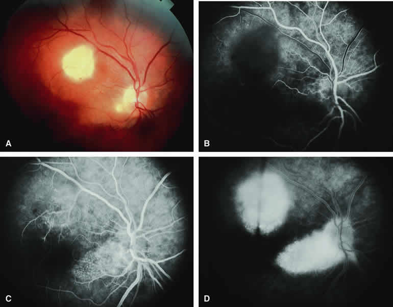
CHOROIDAL METASTATIC CARCINOMA

The typical metastatic carcinoma to the choroid is a yellow, placoid choroidal tumor (Figs. 16A and 17A) associated with shallow subretinal fluid out of proportion to the size of the lesion. Atypical metastatic tumors to the choroid include some cutaneous melanomas, which tend to appear golden brown to dark brown (Fig. 18A), carcinomas that have prominent intralesional blood vessels and, therefore, appear red to orange (Fig. 19A), and rare instances in which a metastatic carcinoma focally erupts through Bruch's membrane to simulate a choroidal melanoma (Fig. 20A). The fluorescein and ICG angiographic features of metastatic carcinomas to the choroid have been described by multiple authors.1, 2, 3, 4, 5, 6, 7, 22, 23

Fig. 16. Typical amelanotic metastatic carcinoma to choroid. A. Yellow-white choroidal tumor located interotemporal from optic disc. Note associated turbid subretinal fluid (most evident in maculopapillary bundle region). B-D. Fluorescein angiogram of lesion. B. Laminar venous phase frame showing ill-defined mottled hypofluorescence and hyperfluorescence corresponding to clinically visible lesion. C. Later laminar venous phase frame still showing no distinct choroidal mass. D. Late-phase frame showing diffuse hyperfluorescence corresponding to lesion and leakage of fluorescein into subretinal space.

Fig. 17. Metastatic carcinoma to choroid with prominent overlying serous subretinal fluid. A. Yellow-white juxtapapillary choroidal tumor with associated secondary retinal detachment. B-D. Fluorescein angiogram of lesion. B. Venous phase frame showing multiple intensely hyperfluorescent smudgy foci at retinal pigment epithelial level overlying ill-defined choroidal mass. C. Later venous phase frame showing generalized hyperfluorescence of mass, increased smudginess of hyperfluorescent foci overlying lesion, and abrupt demarcation between hyperfluorescent choroidal mass and adjacent normal choroid inferiorly. D. Later phase frame showing generalized hyperfluorescence of mass and overlying subretinal fluid.

Fig. 18. Melanotic metastatic cutaneous melanoma to choroid and retina. A. Melanotic metastatic lesions to choroid (three lesions deep to retinal vasculature at left side of photograph) and retina (arcuate lesion in papillomacular distribution). B-D. Fluorescein angiogram of lesions. B. Laminar venous phase frame showing complete hypofluorescence of retinal lesion and patchy partial hypofluorescence corresponding to three choroidal lesions. C. Full venous phase frame showing persistent well-defined hypofluorescence of retinal lesion, slightly increased choroidal hypofluorescence corresponding to the two larger choroidal lesions, and almost no abnormality corresponding to the smallest choroidal lesion. D. Late-phase frame showing persistent nonfluorescence of the retinal lesion but no abnormality corresponding to the three choroidal lesions. (Courtesy of Michael Bourne, MD)

Fig. 19. Follicular thyroid carcinoma metastatic to choroid. A. Reddish orange macular choroidal tumor with surrounding and dependent subretinal blood. B-D. Fluorescein angiogram of lesion. B. Arterial phase frame showing early filling of ill-defined vascular channels within choroidal lesion. C. Later laminar venous phase frame showing intense generalized hyperfluorescence of mass. D. Late-phase frame showing homogeneous intense hyperfluorescence of choroidal mass and choroidal fluorescence blockage corresponding to the surrounding subretinal blood.

Fig. 20. Metastatic lung carcinoma to choroid exhibiting apical nodular eruption of tumor through Bruch's membrane. A. Yellow-white choroidal mass with tan apical nodular eruption through Bruch's membrane, blots of subretinal blood on the surface of the nodule, and an associated secondary retinal detachment. B-D. Fluorescein angiogram of lesion. B. Venous phase frame showing generalized hypofluorescence of choroidal mass except for focal deep intralesional smudgy hyperfluorescent figures. C. Late venous phase frame showing sustained hypofluorescence of choroidal mass, increased hyperfluorescence of smudgy intralesional foci, and hyperfluorescent subretinal fluid along the superior margin of the mass. D. Late-phase frame showing persistent hypofluorescence of most of the apical nodule, expanded smudgy hyperfluorescence of several foci within the lesion, and intense staining of the associated subretinal fluid.

Typical Amelanotic Choroidal Metastatic Tumor

On fluorescein angiography, the typical metastatic tumor (see Figs. 16B, C, and D and 17B, C, and D) appears relatively hypofluorescent in the early frames of the study but becomes progressively more fluorescent as the study continues. Few, if any, intralesional blood vessels are demonstrable. Because of the damaging effects of the expanding choroidal tumor on the overlying retinal pigment epithelium, diffuse or multifocal hypofluorescence and hyperfluorescence are often noted at the RPE level overlying the lesion. In addition, pinpoint hyperfluorescent foci at the RPE level, which are generally attributed to microcystic RPE degeneration, commonly become apparent over the surface of the tumor by the late frames. Fluorescein typically leaks through the retinal pigment epithelium to accumulate in the overlying and surrounding subretinal space by the late frames.

ICG angiography of a typical metastatic choroidal carcinoma23 reveals generalized hypofluorescence of the mass in the early frames, absence of any large-caliber intralesional blood vessels, and mild late hyperfluorescence of the entire lesion. In some lesions, smudgy large-caliber choroidal blood vessels passing through or superficial to the mass can be visualized during the early-to midphase frames of the angiogram. As with other choroidal tumors, ICG angiography seems to show the full basal extent of the tumor substantially better than does fluorescein angiography. In some patients, it even reveals small metastatic choroidal lesions that are not evident by ophthalmoscopy or fluorescein angiography.

Melanotic Metastatic Choroidal Tumor (Metastatic Cutaneous Melanoma)

Occasional patients with metastasis from a primary cutaneous melanoma develop metastatic tumors in the choroid. If the metastatic choroidal lesions are darkly melanotic (see Fig. 18A), they will appear intensely hypofluorescent both early and late on fluorescence angiography (see Fig. 18B, C, and D). In contrast, if the lesions are relatively hypomelanotic, they may exhibit only mild relative hypofluorescence early. Most hypomelanotic metastatic melanomas to the choroid appear relatively hyperfluorescent on late frames of fluorescein angiograms because of progressive fluorescein leakage during the course of the study.

Vascularized Metastatic Choroidal Tumor

Some metastatic carcinomas to the choroid, including follicular thyroid carcinoma, renal cell carcinoma, and particularly choriocarcinoma, have been noted to be associated with prominent intralesional blood vessels that are apparently stimulated to develop by vasogenic humoral factors secreted by the tumor. These tumors tend to have a red to orange color and often give rise to spontaneous subretinal hemorrhages. An example of such a metastatic choroidal tumor is shown in Figure 19A. Fluorescence angiography of this tumor (see Fig. 19B to D) reveals an intralesional vascular network similar to that observed in many hypomelanotic melanomas. The tumor characteristically exhibits early filling of the intralesional blood vessels, intense fluorescence of the entire lesion shortly after initial vascular filling, and intense late staining with leakage of dye into the subretinal fluid. Consequently, detection of this intralesional vascular network does not rule out a metastatic carcinoma to the choroid.

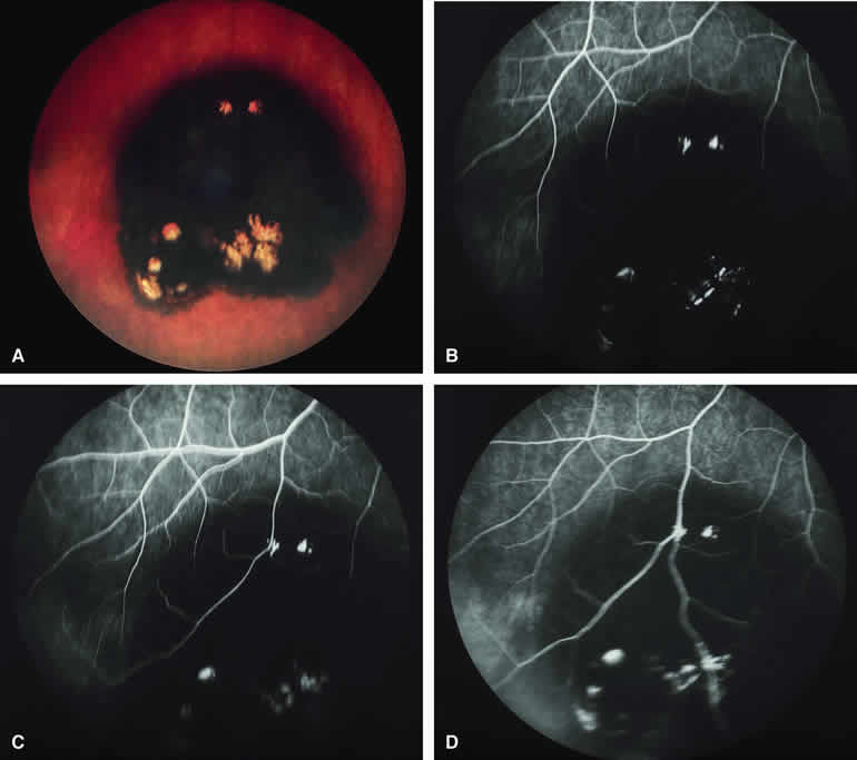
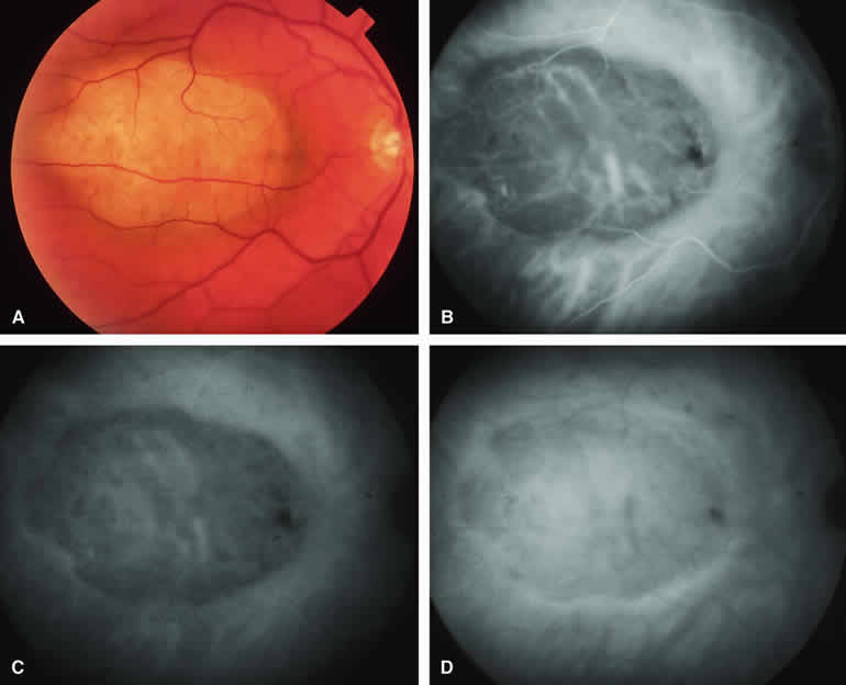
Metastatic Carcinoma to Choroid with Apical Eruption Through Bruch's Membrane

The mushroom-like cross-sectional shape of a choroidal tumor (resulting from focal nodular eruption of the tumor through Bruch's membrane) is such a common feature of choroidal melanoma that many ophthalmologists believe it to be pathognomonic of this neoplasm. Despite this belief, occasional metastatic carcinomas also exhibit this morphologic pattern. Not surprisingly, these metastatic tumors are almost always mistaken clinically for choroidal melanomas. An example of a metastatic choroidal tumor with a nodular eruption through Bruch's membrane is shown in Figure 20A. In contrast with a choroidal melanoma with this feature, a metastatic choroidal tumor with nodular apical extension through Bruch's membrane usually has few if any prominent intralesional blood vessels within the nodule. On fluorescence angiography (see Fig. 20B, C, and D), the apical nodule of a metastatic choroidal tumor is much more likely to exhibit generalized hypofluorescence and much less likely to exhibit fluorescent large-caliber intralesional blood vessels than a similar nodule associated with a choroidal melanoma.

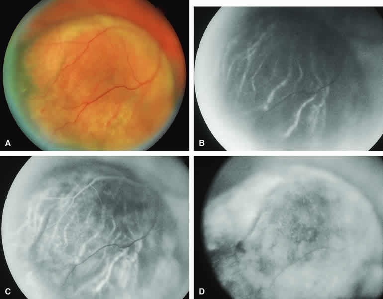
CIRCUMSCRIBED CHOROIDAL HEMANGIOMA

The circumscribed choroidal hemangioma is a benign neoplasm versus hamartoma composed of mature choroidal vascular channels. Fluorescein and ICG angiographic features of these tumors have been described by multiple authors.21,22 The typical circumscribed choroidal hemangioma (Figs. 21A and 22A) is a reddish orange, extremely ill-defined, posterior choroidal tumor that is 5 mm or less in basal diameter and 2 to 3 mm or less in thickness. The overlying retina commonly appears cystic by fundus biomicroscopy, and the retina is often noted to be shallowly detached overlying and surrounding the mass. The posterior margin of virtually all circumscribed choroidal hemangiomas is located within two disc diameters from the optic disc or foveola or both.

Fig. 21. Circumscribed choroidal hemangioma. A. Ill-defined reddish choroidal lesion just superior to optic disc. B-D. Fluorescein angiogram of lesion. B. Laminar venous phase frame showing patchy hyperfluorescence and hypofluorescence of lesion. C. Full venous phase frame showing generalized hyperfluorescence of choroidal tumor with hypofluorescent foci resulting from retinal pigment epithelial (RPE) clumping on surface of lesion. D. Late-phase frame showing persistent generalized hyperfluorescence of lesion.

Fig. 22. Circumscribed choroidal hemangioma. A. Ill-defined choroidal mass just superotemporal to optic disc with central pale zone corresponding to limited fibrous metaplasia of overlying retinal pigment epithelium. B-D. Indocyanine green (ICG) angiogram of lesion. B. Early-phase frame showing generalized hyperfluorescence of choroidal tumor. Note that basal extent of mass is much more apparent on angiogram than on color fundus photograph. C. Intermediate-phase frame showing persistent hyperfluorescence of choroidal mass. D. Very late phase (30 minutes postinjection) frame showing central washout of fluorescein with sustained marginal hyperfluorescence.

Fluorescein angiography of a typical circumscribed choroidal hemangioma (see Fig. 21B, C, and D) shows prominent fluorescence of multiple large-caliber intralesional vessels concurrent with or even before the earliest filling of the normal choroidal and retinal blood vessels. By the late retinal arterial phase frames, the entire lesion is usually diffusely and intensely hyperfluorescent without identifiable distinct intralesional vessels. Fluorescein leaks through the characteristically degenerated overlying retina pigment epithelium into the subretinal space and commonly stains the subretinal fluid and retina diffusely in the late-phase frames.

ICG angiography of a typical circumscribed choroidal hemangioma (see Fig. 22B, C, and D) reveals rapid filling of large-caliber intralesional blood vessels, early intense fluorescence of the vascular tumor, persistent hyperfluorescence of the tumor for 10 to 15 minutes or longer, and late washout of central fluorescence with residual marginal fluorescence by the 20- to 30-minute postinjection frames. This ICG angiographic pattern appears to be almost pathognomonic of circumscribed choroidal hemangiomas and is, therefore, of substantial differential diagnostic value. ICG angiography usually reveals the full extent of the base of the tumor much better and more completely than does fluorescein angiography.

Circumscribed Choroidal Hemangioma with Overlying Fibrous Metaplasia of Retinal Pigment Epithelium

Some circumscribed choroidal hemangiomas exhibit prominent fibrous metaplasia of the retinal pigment epithelium overlying much of the lesion (Fig. 23A). The fibrous metaplasia appears as a white layer of connective tissue deep to the cystic sensory retina overlying the tumor. Fluorescein angiography of these tumors (see Fig. 23B, C, and D) often shows blockage of much of the early vascular filling. In contrast, ICG angiography generally shows the characteristic pattern of intense early hyperfluorescence and late central washout despite the fibrous tissue on the surface of the tumor.
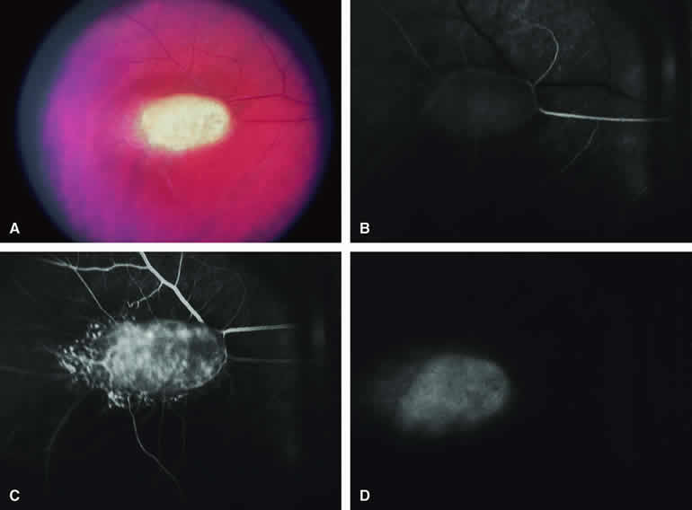
Fig. 23. Circumscribed choroidal hemangioma with prominent overlying fibrous metaplasia of retinal pigment epithelium. A. Nodular reddish pink choroidal mass with overlying whitish fibrous metaplasia of retinal pigment epithelium. B-D. Fluorescein angiogram of lesion. B. Choroidal arterial phase frame showing generalized smudgy vascular fluorescence of choroidal mass. C. Laminar venous phase frame showing intense generalized hyperfluorescence of choroidal mass. D. Full venous phase frame showing sustained generalized hyperfluorescence of choroidal mass.

CHOROIDAL OSTEOMA

The choroidal osteoma is an acquired benign bony tumor of the choroid. It affects women predominantly and usually becomes apparent by the second to third decades of life. The lesion can impair vision substantially but has no malignant potential. The typical lesion appears as a yellowish white to yellow-orange, circumpapillary or juxtapapillary, platelike choroidal mass with well-defined margins (Fig. 24A). These lesions occur bilaterally in approximately 20% to 25% of affected people. Ocular ultrasonography and computed tomography (CT) scanning are of great value in confirming the clinical diagnosis. Fluorescein and ICG angiography of these tumors have been reported by several authors.29, 30, 31

Fig. 24. Choroidal osteoma. A. Yellowish white plate-like macular and juxtapapillary choroidal mass with well-defined margins. B, C. Fluorescein angiogram of lesion. B. Venous phase frame showing generalized hyperfluorescence of mass with spidery hypofluorescent intralesional vascular channels above and focal choroidal fluorescence blockage by blot of subretinal blood inferiorly. C. Later venous phase frame showing findings similar to those of the preceding frame. The distinct upper margin of the choroidal lesion is evident on this image.

Typical Choroidal Osteoma

On fluorescein angiography (see Fig. 24B, C, and C), the characteristic lesion shows numerous spidery superficial intralesional blood vessels in the early frames of the study and patchy or diffuse staining of most or all of the lesion in the late frames. This pattern does not appear to be pathognomonic.

ICG angiograms of several choroidal osteomas have been reported.29,30 These lesions exhibited a prominent early pattern of small intralesional blood vessels, some of which were not evident on fluorescein angiography. Although the lesions appeared generally hypofluorescent early, leakage from the intralesional blood vessels resulted in variable late hyperfluorescence. Patchy areas and foci of late hypofluorescence have been attributed to blockage of choroidal fluorescence by the bone or hyperplastic retinal pigment epithelium overlying the tumor. The full extent of the base of the lesion is generally revealed much more clearly by ICG angiography than by fluorescein angiography.

Choroidal Osteoma with Overlying Choroidal Neovascular Membrane

Because choroidal neovascularization is a relatively frequent complication of choroidal osteomas,31 fluorescence angiography may be performed to evaluate the possible presence and extent of this vascular lesion. When present (Fig. 25A), a choroidal neovascular membrane lesion will appear on fluorescein angiography (see Fig. 25B, C, and D) as an abnormal vascular network beneath the retinal pigment epithelium or sensory retina in the early frames of the study and as a smudgy hyperfluorescent lesion showing progressive increase in fluorescence and late staining with leakage in the late frames. If one treats this neovascular membrane by photocoagulation or photodynamic therapy, one can monitor the response of that lesion with a subsequent angiogram of the same type.

Fig. 25. Choroidal osteoma with overlying choroidal neovascular membrane. A. Yellow-orange macular and juxtapapillary choroidal tumor with well-defined superior margin, prominent large-caliber intralesional blood vessels, a central macular black and red subretinal hemorrhagic figure, and bright red blood with turbid subretinal fluid at the left edge of the image. B-D. Fluorescein angiogram of lesion. B. Laminar venous phase frame showing generalized hyperfluorescence of choroidal lesion, variably intense hypofluorescence corresponding to the subretinal blood, and a partially well-defined choroidal neovascular membrane extending temporally from the central macula. C. Late laminar venous phase frame showing increased fluorescence of choroidal neovascular membrane and early smudgy leakage of fluorescein into overlying subretinal fluid. D. Late-phase frame showing persistent relative hyperfluorescence of choroidal mass, intense smudgy subretinal hyperfluorescence from the portion of the choroidal neovascular membrane at the temporal aspect of the macula, and sustained hypofluorescence corresponding to the subretinal blood.

Back to Top
RETINAL TUMORS
Although choroidal tumors are usually better delineated by ICG angiography than by fluorescein angiography, the fluorescein technique is generally far superior for evaluating intrinsically retinal tumors. Consequently, only fluorescein angiographic features of various retinal tumors are mentioned in this section.

RETINOBLASTOMA

Retinoblastoma is the most common primary intraocular malignancy in children. It is usually a unilateral, unifocal, sporadic disease, but a substantial proportion of affected children have multifocal, bilateral, genetic retinoblastoma. A typical discrete retinal tumor in retinoblastoma appears as an opaque white retinal mass with prominent small-caliber surface blood vessels and dilated tortuous afferent and efferent retinal shunt vessels (Figs. 26A and 27A). Unfortunately, most children with retinoblastoma present at a much more advanced intraocular extent of disease, exhibiting leukocoria resulting from the bulk of white tumor filling the vitreous chamber.

Fig. 26. Small intraretinal retinoblastoma. A. Small poorly defined white intraretinal mass with fine intralesional vascular channels. B-D. Fluorescein angiogram of lesion. B. Arterial phase frame showing filling of retinal arteries supplying tumor. C. Later arterial phase frame showing filling of capillary network within tumor. D. Late-phase frame showing homogeneous mild staining of tumor.

Fig. 27. Intraretinal retinoblastoma. A. Well-defined nodular white retinal tumor with prominent retinal vasculature extending to and from lesion. B-D. Fluorescein angiogram of lesion. B. Arterial phase frame showing rapid filling of feeding retinal arteries and capillary network within lesion. C. Laminar venous phase frame showing more complete filling of tumor vasculature. D. Late-phase frame showing intense staining of entire tumor.

Because retinoblastoma usually affects young children, fluorescein angiography must be obtained during examination under anesthesia in most cases. Performing a fluorescein angiogram on an intubated infant or small child requires a fundus camera that is mounted vertically on a mobile base or special positioning of the child.32 Fundus fluorescein angiography is rarely, if ever, helpful in the diagnosis of retinoblastoma. If one can visualize the fundus adequately by ophthalmoscopy and can identify discrete white retinal tumors supplied by and drained by retinal blood vessels, fluorescein angiography provides no essential additional information that establishes or refutes the diagnosis of retinoblastoma. The only true solid tumor that strongly resembles retinoblastoma, the retinal astrocytic hamartoma, usually does not have associated dilated and tortuous retinal blood vessels or the degree of surface vascularity that one expects with retinoblastoma.

Discrete Intraretinal Retinoblastoma

If fluorescein angiography is performed on a discrete intraretinal retinoblastoma (see Figs. 26B, C, and D and 27B, C, and D), the angiogram typically shows rapid filling of the feeding retinal artery, prompt filling of the intralesional capillaries within a second or two, and rapid subsequent filling of the draining retinal vein.32 The intralesional capillaries tend to leak fluorescein, so the tumor characteristically stains brightly in the late frames. However, intraretinal retinoblastoma lesions typically do not show substantial fluorescein leakage into the overlying vitreous.

Larger intraretinal retinoblastomas are typically associated with multiple dilated and tortuous feeding retinal arteries and draining retinal veins (Fig. 28A). Fluorescein angiography of this tumor (see Fig. 28B, C, and D) shows rapid filling of the feeding arteries, rapid subsequent filling of intralesional blood vessels, and relatively quick filling of all draining retinal veins. In the later frames, it exhibits intense hyperfluorescent staining.

Fig. 28. Larger intraretinal retinoblastoma with prominent retinal vasculature. A. Nodular white retinal tumor with associated dilated tortuous feeding retinal arteries and draining retinal veins. B-D. Fluorescein angiogram of lesions. B. Arterial phase frame showing rapid filling of retinal arteries feeding tumor. C. Early laminar venous phase frame showing fluorescence of large-caliber intralesional blood vessels. D. Late-phase frame showing diffuse hyperfluorescence of entire tumor and marked tortuosity of draining retinal veins.

Advanced Intraocular Retinoblastoma Presenting as Leukocoria

In cases of total leukocoria in which one cannot discern a distinct tumor, fluorescein angiography documents the visible vascular abnormalities in the detached retrolental retina (exophytic growth pattern) or the absence of vessels in the fluffy retrolental tumor (endophytic growth pattern) but is not otherwise of diagnostic value.

Regressed Intraretinal Retinoblastoma Following Radiation Therapy

If retinoblastoma is managed by radiation therapy, the tumors commonly regress to calcified (type I) residual nonviable lesions. In some cases, however, a substantial fish flesh (type II) regression pattern accompanies a partially calcific response. Although the entire tumor or portion that exhibits this regression pattern typically appears more grayish and less vascular than untreated viable tumors, this distinction is occasionally difficult to make by ophthalmoscopy. In these situations, fluorescein angiography may be helpful in distinguishing between residual viable tumor and satisfactory type II regression. If the tumor is regressed in a type II pattern, fluorescein angiography typically shows the afferent and efferent retinal vessels to be much less prominent than they were before treatment, the residual intralesional vasculature to be markedly diminished or absent, and late staining to be mild.

Regressed Retinoblastoma Following Chemotherapy

The first-line management of many children with retinoblastoma today is chemotherapy rather than radiation therapy. Retinoblastomas that have regressed completely following chemotherapy exhibit few if any residual patent intralesional blood vessels and limited late hyperfluorescence. In contrast, retinoblastomas that remain viable and likely to relapse locally tend to have some persistent intralesional blood vessels that can show up prominently on fluorescein angiography.

Regressed Retinoblastoma Following Photocoagulation or Laser Therapy

Following successful photocoagulation or laser therapy of localized retinoblastoma, the entire treated site should appear flat and atrophic. If there is any question about residual tumor, fluorescein angiography may be helpful. If the lesion is indeed fully regressed, there should be no residual abnormal vasculature within the central portion of the lesion. If even a small amount of residual viable tumor is present, one will often note an irregular vasculature corresponding to that lesion and late staining of that portion. A fluorescein angiogram showing these features is probably an indication for supplemental tumor treatment.

RETINAL ASTROCYTIC HAMARTOMA

The retinal astrocytic hamartomas is a benign neoplasm derived from intraretinal astrocytes. The typical lesion becomes evident in childhood or early adulthood. This lesion is a frequent feature of tuberous sclerosis, but some patients develop an astrocytic hamartoma of the retina in the absence of other features of this syndrome.

Typical Retinal Astrocytic Hamartomas in Tuberous Sclerosis

Typical retinal astrocytic hamartomas associated with tuberous sclerosis appear as translucent patches, opalescent spots, or opaque white retinal tumors33,34 (Figs. 29A and 30A). Affected people often have more than one lesion in the affected eye, bilateral involvement, or both. Fluorescein angiography of these lesions (see Figs. 29B, C, and D and 30B, C, and D) tends to show relatively normal filling of the retinal arteries leading to the tumor, a limited intralesional vascular network, normal slow filling of the retinal veins draining the lesion, and mild late staining of the tumors.

Fig. 29. Retinal astrocytic hamartoma in child with tuberous sclerosis. A. Unifocal white inner retinal tumor without associated dilation or tortuosity of adjacent retinal arteries and veins. B-D. Fluorescein angiogram of lesion. B. Arterial phase frame showing faint pseudofluorescence of tumor and limited early filling of intralesional vasculature. C. Venous phase frame showing fluorescence of irregular capillary network within lesion. D. Late-phase frame showing staining of tumor.

Fig. 30. Multifocal retinal astrocytic hamartomas in tuberous sclerosis. A. Two white intraretinal lesions (larger lesion in superior macula, smaller lesion at inferotemporal margin of optic disc) in young adult patient with tuberous sclerosis. B-D. Fluorescein angiogram of lesions. B. Laminar venous phase fluorescein angiogram showing hypofluorescence of superior macular and inferotemporal maculopapillary bundle lesions. C. Venous phase frame showing smudgy hyperfluorescence corresponding to both retinal lesions. D. Late-phase frame showing diffuse staining of both lesions.

Isolated Retinal Astrocytic Hamartoma

The isolated retinal astrocytic hamartoma35 (Fig. 31A) is a unilateral, unifocal, pale yellow to dull white fundus lesion that is usually mistaken for an amelanotic melanoma with a broad area of retinal invasion at its apex. These lesions tend to occur in teenagers to middle-aged adults. They tend to enlarge over time and are usually complicated by a progressive exudative retinal detachment that causes secondary visual loss. Fluorescein angiography of this tumor (see Fig. 31B, C, and D) typically reveals a prominent irregular retinal vascular network on the surface of the tumor and intense late fluorescence of the mass with leakage of dye into the associated subretinal fluid.

Fig. 31. Isolated retinal astrocytic hamartoma in patient with no evidence of tuberous sclerosis. A. Pale yellow macular fundus lesion overlying optic disc associated with bullous nonrhegmatogenous retinal detachment. B-D. Fluorescein angiogram of lesion. B. Arterial phase frame showing hypofluorescence of nasal aspect of lesion and unusual pattern of retinal blood vessels on surface of mass. C. Venous phase frame showing prominent retinal capillary network within mass. D. Late-phase frame showing intense late staining of mass and associated subretinal fluid.

RETINAL CAPILLARY HEMANGIOMA (VON HIPPEL TUMOR)

The typical retinal capillary hemangioma is distinctive in its ophthalmoscopic appearance. The lesion is characteristically red-orange and associated with prominent afferent and efferent retinal vessels (Figs. 32A and 33A). Fluorescein angiography of these tumors has been described by multiple authors.1, 2, 3, 36, 37

Fig. 32. Small retinal capillary hemangioma. A. Spherical red vascular lesion in superotemporal periphery associated with prominent dilated, tortuous afferent and efferent retinal vascular channels. B-D. Fluorescein angiogram of lesion. B. Arterial phase frame showing filling of afferent arterial channel and lesion. C. Venous phase frame showing intense hyperfluorescence of vascular lesion, as well as complete filling of both afferent and efferent channels. D. Late-phase frame showing smudgy hyperfluorescence of lesion resulting from leakage of fluorescein into overlying vitreous and surrounding retina. Hypofluorescent and hyperfluorescent lesion at top of figure is site of a previously coagulated retinal capillary hemangioma.

Fig. 33. Larger retinal capillary hemangioma. A. Spherical pale red retinal lesion associated with dilated tortuous retinal arteries and veins and macular subretinal exudates. B-D. Fluorescein angiogram of lesion. B. Arterial phase frame showing dilation and tortuosity of feeding retinal artery and early hyperfluorescent filling of the vascular lesion. C. Venous phase frame showing intense hyperfluorescence of entire lesion plus fluorescein filling of the previously unfilled draining retinal vein. D. Late-phase frame showing smudgy leakage of fluorescein from the persistently hyperfluorescent retinal vascular lesion.

If fluorescein angiography is performed on a typical retinal capillary hemangioma (see Figs. 32B, C, and D and 33B, C, and D), it shows rapid filling of the afferent artery, brisk filling of the entire retinal vascular lesion, and intense hyperfluorescence of the entire vascular lesion shortly thereafter, followed by rapid filling of the efferent vessel. Retinal capillary hemangiomas characteristically leak fluorescein exuberantly into the overlying vitreous, so that the lesion's margins appear fuzzy in the late-phase frames. Because many retinal capillary hemangiomas are also associated with exudative subretinal fluid, the fluorescein commonly accumulates in the subretinal space by the late frames.

In patients with a relatively large peripheral retinal capillary hemangioma (Fig. 34A), the retinal arteries leading from the optic disc to the tumor and the retinal veins leading back from the tumor to the optic disc typically appear enormously dilated and markedly tortuous. On fluorescein angiography (see Fig. 34B, C, and D), the feeding artery tends to fill rapidly and the draining retinal vein similarly fills shortly thereafter. The peripheral retinal capillary hemangioma tends to become intensely hyperfluorescent with profuse leakage of fluorescein into the adjacent vitreous.

Fig. 34. Relatively large peripheral retinal capillary hemangioma associated with subtotal tractional-exudative retinal detachment. A. Prominent red to white peripheral fundus lesion superotemporally associated with tractional-exudative retinal detachment. B-D. Fluorescein angiogram of lesion. B. Early laminar venous phase frame of posterior pole showing massive dilation and tortuosity of fluorescein-filled superotemporal branch retinal artery and unfilled superotemporal branch retinal vein. C. Venous phase frame of posterior pole showing fluorescein filling of both the superotemporal branch retinal artery and vein. D. Venous phase frame showing diffusely hyperfluorescent peripheral retinal lesion.

Fluorescein angiography may help in detection of extremely subtle retinal capillary hemangiomas not identifiable by ophthalmoscopy alone. To detect these lesions, one has to be sure that the angiogram adequately surveys all quadrants of the fundus from the posterior pole to the periphery (as far as can be visualized photographically). Subtle small retinal capillary hemangiomas need not necessarily be treated, but eyes with these lesions should certainly be watched closely for any lesion progression that might eventually warrant treatment.

RETINAL CAVERNOUS HEMANGIOMA

The cavernous hemangioma of the retina is a benign retinal vascular lesion that has characteristic clinical and angiographic features. The typical cavernous hemangioma of the retina (Fig. 35A) appears as a small cluster of intraretinal vascular saccules that can appear similar to localized retinal telangiectasia in a limited form of Coats' disease. Fluorescein angiographic features of retinal cavernous hemangiomas have been described by several authors.2,3,38,39

Fig. 35. Retinal cavernous hemangioma. A. Cluster of dark red vascular saccules just superotemporal from center of macula along course of superotemporal branch retinal vein. White tissue on surface of lesion corresponds pathologically to gliosis. B-D. Fluorescein angiogram of lesion. B. Laminar venous phase angiogram showing abnormal retinal venous channels passing through largely hypofluorescent lesion. C. Full venous phase frame showing filling in of many vascular saccules comprising lesion. D. Late-phase frame showing hyperfluorescent cap of many component saccules of lesion, attributable to plasma-erythrocyte separation.

Fluorescein angiography of a typical cavernous hemangioma of the retina (see Fig. 35B, C, and D) shows a hypofluorescent retinal zone corresponding to the area of the lesion in the early-phase frames; an anomalous draining retinal vein passing through the center of the lesion; and slow progressive filling of the vascular saccules comprising the lesion, usually from the periphery of the lesion toward its center. The vascular saccules generally do not leak, and there is no evidence of associated broadening of the intercapillary spaces or retinal capillary nonperfusion peripherally. In the late frames, one commonly observes the phenomenon of plasma-erythrocyte separation in which the red blood cells gravitate to the dependent portion of individual vascular saccules while the relatively acellular plasma remains above. The plasma component of the saccules tends to stain intensely with fluorescein in the late frames, whereas the dependent, erythrocyte-filled portion remains intensely hypofluorescent.

COMBINED HAMARTOMA OF RETINA

The combined hamartoma of the retina is an uncommon congenital benign lesion of the sensory retina, retinal pigment epithelium, and retinal vasculature. The typical lesion occurs in a juxtapapillary or circumpapillary location and appears as a shaggy gray and white full-thickness retinal lesion with pronounced tortuosity of the incorporated retinal blood vessels and prominent surface fibrosis (Fig. 36A). The retinal blood vessels beyond the lesion appear unremarkable in caliber and course, and the overlying vitreous is clear. Because of the characteristic appearance of these lesions, fluorescein angiography40 is usually of documentary rather than of diagnostic importance.

Fig. 36. Combined hamartoma of retina. A. Ill-defined gray circumpapillary retinal lesion with whitish gliosis overlying portion of optic disc and prominent vascular tortuosity within lesion. B-D. Fluorescein angiogram of lesion. B. Arterial phase frame showing prominent tortuosity of large-caliber retinal blood vessels involved in mass. C. Laminar venous phase frame showing similar tortuosity of retinal veins and venules within lesion. D. Full venous phase frame showing ill-defined hypofluorescent background corresponding to darkly pigmented portion of mass.

The fluorescein angiogram of a typical combined retinal hamartoma (see Fig. 36B, C, and D) shows early hypofluorescence corresponding to the lesion (attributable to the associated RPE proliferation), intralesional retinal vascular tortuosity, gradual leakage of fluorescein from the tortuous intralesional vessels, and diffuse late staining of the entire lesion. The retinal blood vessels beyond the margins of the lesion tend to be normal in course and caliber and do not leak. The retinal pigment epithelium around the margin of the lesion occasionally shows radiating hyperfluorescent striae attributable to RPE depigmentation.

UNIFOCAL HYPERTROPHY OF RETINAL PIGMENT EPITHELIUM

The unifocal hypertrophy of the retinal pigment epithelium is a benign congenital lesion of extremely limited malignant potential. The typical lesion is a flat to minimally thick, gray to black, well-defined, nummular lesion up to 5 mm in diameter (Fig. 37A). The overlying large-caliber retinal vessels usually appear normal by fundus biomicroscopy. Because of the characteristic appearance of these lesions, fluorescein angiography is not needed for diagnosis. If one performs fluorescein angiography of this lesion (see Fig. 37B, C, and D), however, one expects the entire lesion to be hypofluorescent throughout the entire study. The overlying retinal vasculature tends to be normal in most cases.41

Fig. 37. Congenital hypertrophy of retinal pigment epithelium. A. Flat gray to black retinal pigment epithelial level lesion with well-defined smooth margins. This lesion exhibits partial depigmentation peripherally. B-D. Fluorescein angiogram of lesion. B. Arterial phase frame showing hypofluorescence corresponding to lesion. Note faint transmission hyperfluorescence resulting from thinning of retinal pigment epithelium in marginal zone of lesion. C. Laminar venous phase frame showing most of lesion to be persistently hypofluorescent; however, zones of transmission hyperfluorescence are more apparent. D. Late-phase frame showing persistent hypofluorescence of lesion and fading of transmission hyperfluorescence.

Variations on this normal pattern occur in unifocal hypertrophy lesions that develop foci of depigmentation (lacunae) (Fig. 38A). On fluorescein angiography (see Fig. 38B, C, and D), these foci appear as window defects of hyperfluorescence that appear early in the study and persist in the late frames without change in their margins.

Fig. 38. Congenital hypertrophy of retinal pigment epithelium with prominent lacunae of depigmentation. A. Well-defined black peripheral fundus lesion with several discrete foci of almost complete depigmentation (lacunae). B-D. Fluorescein angiogram of lesion. B. Venous phase frame showing well-defined choroidal fluorescence blockage corresponding to lesion and hyperfluorescent foci corresponding to depigmented lacunae. C. Later venous phase frame showing features similar to those on the prior image. D. Recirculation phase frame showing persistent hypofluorescence of lesion and sustained well-defined hyperfluorescence of focal lacunae.

PRIMARY INTRAOCULAR LYMPHOMA

Primary intraocular lymphoma is a rare ophthalmic malignancy that occurs in two principal forms. Primary vitreoretinal lymphoma42,43 is the more common of these two forms. It generally develops in older adult patients. The usual presenting symptoms are floaters and blurred vision in one or both eyes. Classic ocular findings in this form of lymphoma include diffuse intravitreal pale cells and geographic subretinal pigment epithelial accumulations of tumor cells (Fig. 39A). The sub-RPE lesions may be unifocal or multifocal and are often bilateral. Occasional patients with this form of lymphoma will develop discrete intraretinal infiltrates of tumor cells, often in association with intraretinal and intravitreal bleeding. This vitreoretinal form of lymphoma is strongly associated with antecedent, concurrent, or subsequent primary CNS lymphoma.

Fig. 39. Lymphomatous subretinal pigment epithelial infiltrate. A. Geographic yellow-white subretinal pigment epithelial lesion in temporal midzone. Note smaller satellite lesions and clumped retinal pigment epithelial pigment on surface of large lesion. B-D. Fluorescein angiogram of lesion. B. Venous phase frame showing hypofluorescence corresponding to geographic fundus lesion and satellite lesions. C. Recirculation phase frame showing marginal hyperfluorescence of geographic lesion and smudgy hyperfluorescence of satellite lesions. D. Late-phase frame showing mild hyperfluorescence of geographic lesion and intense hyperfluorescence of satellite lesions.

The second classic type of primary intraocular lymphoma is the diffuse uveal lymphoma.44,45 This form of the disease appears as diffuse thickening of the uveal tract, sometimes with accentuated focal nodules. The involved choroid appears creamy, and the overlying retinal pigment epithelium is often disrupted with patches of RPE hyperplasia. In rare cases, an unusual reticular accumulation of lipofuscin pigment develops on the surface of the thickened choroid, giving the fundus an appearance that resembles fundus flavimaculatus. This diffuse uveal form of primary intraocular lymphoma tends to be strongly associated with antecedent, concurrent, or subsequent visceral non-Hodgkin's lymphoma.

Geographic Subretinal Pigment Epithelial Lesions

The subretinal pigment epithelial lesions that are characteristic of the vitreal-central nervous system form of diffuse large cell lymphoma (reticulum cell sarcoma) have a characteristic ophthalmoscopic appearance, which is readily recognized in most cases. The classic lesion is a geographic yellow-white mass deep to the retina and retinal pigment epithelium associated with smaller and less well-defined satellite lesions at the same level (see Fig. 39A). The retinal pigment epithelium overlying these lesions is partially disrupted, and black pigment clumps are evident on the surface of these tumors. Fluorescein angiography is unlikely to establish the diagnosis if this disorder is not suspected clinically.

Fluorescein angiography of a typical subretinal pigment epithelial infiltrate (see Fig. 39B, C, and D) shows the geographic lesion to be hypofluorescent during the early-phase frames with gradual marginal hyperfluorescence developing as the study continues to the late frames. There is rarely any significant abnormality of the overlying retinal vasculature or prominent late leakage of fluorescein into the subretinal space.

Lymphomatous Retinal Infiltrates

When primary vitreoretinal lymphoma appears as a retinal infiltrate,43 fluorescein angiography typically shows blockage of choroidal and retinal fluorescence by the lesion in the early-phase frames, irregular leaking retinal blood vessels at the margins of the lesion, and progressive leakage of fluorescein from the retinal vessels into the lesion and overlying vitreous. This angiographic pattern does not clearly distinguish these lesions from microbial retinal lesions that they frequent simulate.

METASTATIC CARCINOMA TO RETINA

Although the choroid is the predominant intraocular site for metastatic carcinomas, the retina is occasionally involved as a metastatic site.46 Metastatic retinal tumors generally appear as pale yellow to white retinal infiltrates, which may be unifocal or multifocal and are often associated with vitreous cells.

Amelanotic Metastasis to Retina

An amelanotic metastasis to the retina appears as an ill-defined pale retinal infiltrate that obscures at least some of the retinal blood vessels (Fig. 40A). These lesions are often associated with patches of intraretinal blood and occasionally retinal exudates. Fluorescein angiography of such lesions (see Fig. 40B, C, and D) reveals mild hypofluorescence of the lesion in the early frames, multiple irregular small-caliber retinal blood vessels within the lesion, and profuse late leakage and hyperfluorescence of the lesion.

Fig. 40. Amelanotic metastatic carcinoma to retina. A. Pale gray retinal infiltrate at temporal margin of photograph with associated intraretinal hemorrhages and hard exudates. The primary lesion was a cutaneous malignant melanoma. B-D. Fluorescein angiogram of lesion. B. Laminar venous phase frame showing abnormal retinal capillary network corresponding to tumor and fluorescence blockage corresponding to the retinal hemorrhages. C. Full venous phase frame showing intense smudgy hyperfluorescence of neoplastic retinal infiltrate. D. Late-phase frame showing intense expanded hyperfluorescence of the retinal lesion, diffuse hyperfluorescence of the surrounding retina and subretinal fluid, and persistent fluorescence blockage by the intraretinal blood.

Melanotic Metastasis to Retina (Metastatic Cutaneous Melanoma)

A darkly melanotic metastasis to the retina appears as a well-defined dark brown intraretinal lesion (see Fig. 18A). On fluorescein angiography (see Fig. 18B, C, and D), the melanotic retinal infiltrate blocks both choroidal and retinal fluorescence throughout virtually all of the study. The retinal blood vessels around the margins of the metastatic lesion may leak fluorescein by the late-phase frames.

Back to Top
SUMMARY
Fluorescence angiography is an appropriate documentary study for assorted choroidal and retinal tumors. Unfortunately, it is rarely diagnostic in its own right, and the findings identified on fluorescence angiography must always be interpreted in light of the ophthalmoscopic appearance of the lesion and other clinical information. Fluorescence angiography provides limited, if any, clinically significant prognostic information in patients with a choroidal or retinal tumor, but it may be helpful by revealing unanticipated findings inconsistent with the presumptive clinical diagnosis before the angiogram and in identifying inadequate treatment of some tumors. Fluorescence angiography may also be able to document the full extent of a choroidal or retinal tumor more precisely than conventional color fundus photography, thereby enabling the clinician to detect a subtle change in the lesion over time that might otherwise have been overlooked.

Fluorescein and ICG angiography have added more to the understanding of the clinical pathophysiology of choroidal and retinal tumors and the mechanisms for visual loss associated with these lesions than to clarifying diagnosis or prognosis. Even so, fluorescence angiography appears to have a well-established role in the evaluation of patients with retinal and choroidal tumors, and its continued use in the foreseeable future seems appropriate.

Back to Top
REFERENCES

1. Snyder WB, Allen L, Frazier O. Fluorescence angiography of ocular tumors. Trans Am Acad Ophthalmol Otolaryngol 1967;71:820

2. Gass JDM. Fluorescein angiography: An aid in the differential diagnosis of intraocular tumors. Int Ophthalmol Clin 1972;12:85

3. Gass JDM. Differential Diagnosis of Intraocular Tumors: A Stereoscopic Presentation. St Louis: Mosby, 1974

4. Guyer DR, Yannuzzi LA, Krupsky S et al. Digital indocyanine green videoangiography of intraocular tumors. Sem Ophthalmol 1993;8:224

5. Sallet G, Amoaku WM, Lafaut BA et al. Indocyanine green angiography of choroidal tumors. Graefes Arch Clin Exp Ophthalmol 1995;233:677

6. Shields CL, Shields JA, De Potter P. Patterns of indocyanine green videoangiography of choroidal tumors. Br J Ophthalmol 1995;79:237

7. Schalenbourg A, Zografos L, Chamot L et al. Value of indocyanine green video-angiography in the differential diagnosis of non-pigmented choroidal tumors. Klin Monatsbl Augenheilkd 1996;208:330

8. Meyer K, Augsburger JJ. Independent diagnostic value of fluorescein angiography in the evaluation of intraocular tumors. Graefes Arch Clin Exp Ophthalmol 1999;237:489

9. Shiraki K, Moriwaki M, Yanagihara N et al. Indocyanine green angiograms of choroidal nevi. Comparison between confocal and nonconfocal scanning laser ophthalmoscope and fundus video camera. Jpn J Ophthalmol 2001;45:368

10. Andersen MV, Schrfig E, Prause JU. Differential diagnosis of choroidal melanomas and nevi using scanning laser ophthalmoscopical indocyanine green angiography. Acta Ophthalmol 1995;73:453

11. Pro M, Shields JA, Tomer TL. Serous detachment of the macula associated with presumed choroidal nevi. Arch Ophthalmol 1978;96:1374

12. Waltman DD, Gitter KA, Yannuzzi L et al. Choroidal neovascularization associated with choroidal nevi. Am J Ophthalmol 1978;85:704

13. Augsburger JJ, Schroeder RP, Territo C et al. Clinical parameters predictive of enlargement of melanocytic choroidal lesions. Br J Ophthalmol 1989;73:911

14. Edwards WC, Layden WE, MacDonald R. Fluorescein angiography of malignant melanoma of the choroid. Am J Ophthalmol 1969;68:797

15. Petit TH, Barton A, Foos RY et al. Fluorescein angiography of choroidal melanomas. Arch Ophthalmol 1970;83:27

16. Wolter JR, Schut AL, Martonyi CL. Hemangioma-like clinical appearance of collar-button melanoma caused by the strangulation effect of Bruch's membrane. Am J Ophthalmol 1973;76:730

17. Augsburger JJ, Golden MI, Shields JA. Fluorescein angiography of choroidal malignant melanomas with retinal invasion. Retina 1984;4:232

18. Schneider U, Gelisken F, Inhoffen W et al. Indocyanine green videoangiography of malignant melanomas of the choroid using the scanning laser ophthalmoscope. Ger J Ophthalmol 1996;5:6

19. Mueller AJ, Freeman WR, Folberg R et al. Evaluation of microvascularization pattern visibility in human choroidal melanomas: Comparison of confocal fluorescein with indocyanine green angiography. Graefes Arch Clin Exp Ophthalmol 1999;237:448

20. Atmaca LS, Batioglu F, Atmaca P. Fluorescein and indocyanine green videoangiography of choroidal melanomas. Jpn J Ophthalmol 1999;43:25

21. Schaller UC, Mueller AJ, Bartsch DU et al. Choroidal melanoma microcirculation with confocal indocyanine green angiography before and 1 year after radiation brachytherapy. Retina 2000;20:627

22. David DL, Robertson DM. Fluorescein angiography of metastatic choroidal tumors. Arch Ophthalmol 1973;89:97

23. Harino S, Miyamoto K, Okada M et al. Indocyanine green videoangiographic findings in choroidal metastatic tumor. Graefes Arch Clin Exp Ophthalmol 1995;233:339

24. Norton EWD, Gutman F. Fluorescein angiography and hemangiomas of the choroid. Arch Ophthalmol 1967;78:121

25. Bottone F, Trevaert DC, Deutman AF. Fluorescein angiographic findings and results of laser treatment in circumscribed choroidal hemangioma. Int Ophthalmol 1990;14:259

26. Piccolino FC, Borgia L, Zinicola E. Indocyanine green angiography of circumscribed choroidal hemangiomas. Retina 1996;16:19

27. Arevalo JF, Shields CL, Shields JA et al. Circumscribed choroidal hemangioma: Characteristic features with indocyanine green videoangiography. Ophthalmology 2000;107:344

28. Schalenbourg A, Piguet B, Zografos L. Indocyanine green angiographic findings in choroidal hemangiomas: A study of 75 cases. Ophthalmologica 2000;214:246

29. Yuzawa M, Kawamura A, Haruyama M et al. Indocyanine green video angiographic findings in choroidal osteoma. Eur J Ophthalmol 1994;4:191

30. Lafaut BA, Mestdagh C, Kohno T et al. Indocyanine green angiography in choroidal osteoma. Graefes Arch Clin Exp Ophthalmol 1997;235:330

31. Grand MG, Burgess DB, Singerman LJ et al. Choroidal osteoma. Treatment of associated subretinal neovascular membrane. Retina 1984;4:84

32. Ohnishi Y, Yamana Y, Minei M et al. Application of fluorescein angiography in retinoblastoma. Am J Ophthalmol 1982;93:578

33. Nyboer JH, Robertson DM, Gomez MR. Retinal lesions in tuberous sclerosis. Arch Ophthalmol 1976;94:1277

34. Williams R, Taylor D. Tuberous sclerosis. Surv Ophthalmol 1985;30:143

35. Arnold AC, Hepler RS, Yee RW et al. Solitary retinal astrocytoma. Surv Ophthalmol 1985;30:173

36. Gass JDM. Angiomatosis retinae (von Hippel's disease). In Differential Diagnosis of Intraocular Tumors: A Stereoscopic Presentation. St Louis: Mosby, 1974:265–292

37. Augsburger JJ, Shields JA, Goldberg RE. Classification and management of hereditary retinal angiomas. Int Ophthalmol 1981;4:93

38. Colvard DM, Robertson DM, Trautman JC. Cavernous hemangioma of the retina. Arch Ophthalmol 1978;96:2042

39. Messmer E, Laqua H, Wessing A et al. Nine cases of cavernous hemangioma of the retina. Am J Ophthalmol 1983;95:383

40. Schachat AP, Shields JA, Fine SL et al. Combined hamartomas of the retina and retinal pigment epithelium. Ophthalmology 1984;91:1609

41. Cohen SY, Quentel G, Guiberteau B et al. Retinal vascular changes in congenital hypertrophy of the retinal pigment epithelium. Ophthalmology 1993;100:471

42. Gass JDM, Sever RJ, Grizzard WS et al. Multifocal pigment epithelial detachments by reticulum cell sarcoma: A characteristic funduscopic picture. Retina 1984;4:135

43. Ridley ME, McDonald HR, Sternberg P et al. Retinal manifestations of ocular lymphoma (reticulum cell sarcoma). Ophthalmology 1992;99:1153

44. Jakobiec FA, Sacks E, Kronish JW et al. Multifocal static creamy choroidal infiltrates. An early sign of lymphoid neoplasia. Ophthalmology 1987;94:397

45. Gass JDM, Weleber RG, Johnson DR. Non-Hodgkin's lymphoma causing fundus picture simulating fundus flavimaculatus. Retina 1987;7:209

46. Leys AM, Van Eyck LM, Nuttin BJ et al. Metastatic carcinoma to the retina. Clinicopathologic findings in two cases. Arch Ophthalmol 1990;108:1448

Back to Top