|
|
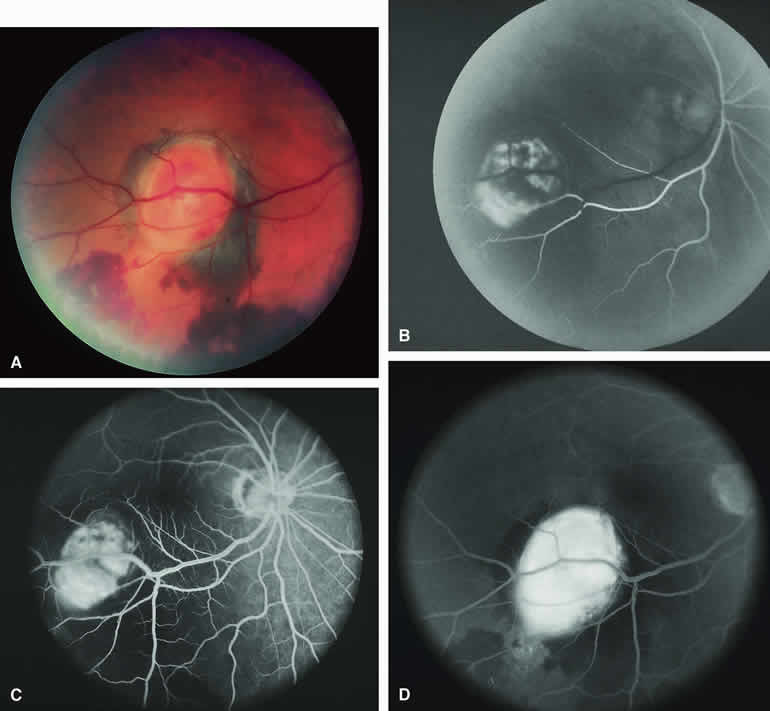
| Fig. 19. Follicular thyroid carcinoma metastatic to choroid. A. Reddish orange macular choroidal tumor with surrounding and dependent subretinal blood. B-D. Fluorescein angiogram of lesion. B. Arterial phase frame showing early filling of ill-defined vascular channels within choroidal lesion. C. Later laminar venous phase frame showing intense generalized hyperfluorescence of mass. D. Late-phase frame showing homogeneous intense hyperfluorescence of choroidal mass and choroidal fluorescence blockage corresponding to the surrounding subretinal blood. |