|
|
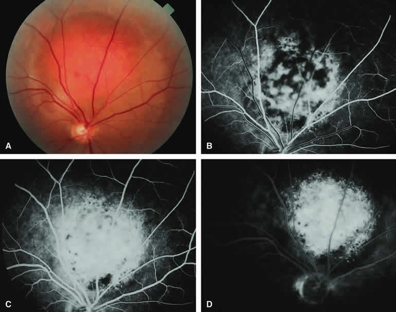
| Fig. 21. Circumscribed choroidal hemangioma. A. Ill-defined reddish choroidal lesion just superior to optic disc. B-D. Fluorescein angiogram of lesion. B. Laminar venous phase frame showing patchy hyperfluorescence and hypofluorescence of lesion. C. Full venous phase frame showing generalized hyperfluorescence of choroidal tumor with hypofluorescent foci resulting from retinal pigment epithelial (RPE) clumping on surface of lesion. D. Late-phase frame showing persistent generalized hyperfluorescence of lesion. |