|
|
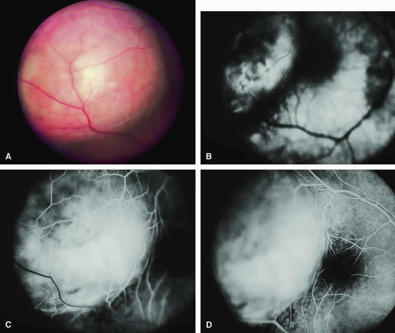
| Fig. 23. Circumscribed choroidal hemangioma with prominent overlying fibrous metaplasia of retinal pigment epithelium. A. Nodular reddish pink choroidal mass with overlying whitish fibrous metaplasia of retinal pigment epithelium. B-D. Fluorescein angiogram of lesion. B. Choroidal arterial phase frame showing generalized smudgy vascular fluorescence of choroidal mass. C. Laminar venous phase frame showing intense generalized hyperfluorescence of choroidal mass. D. Full venous phase frame showing sustained generalized hyperfluorescence of choroidal mass. |