|
|
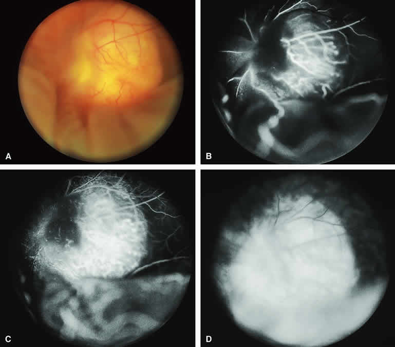
| Fig. 31. Isolated retinal astrocytic hamartoma in patient with no evidence of tuberous sclerosis. A. Pale yellow macular fundus lesion overlying optic disc associated with bullous nonrhegmatogenous retinal detachment. B-D. Fluorescein angiogram of lesion. B. Arterial phase frame showing hypofluorescence of nasal aspect of lesion and unusual pattern of retinal blood vessels on surface of mass. C. Venous phase frame showing prominent retinal capillary network within mass. D. Late-phase frame showing intense late staining of mass and associated subretinal fluid. |