|
|
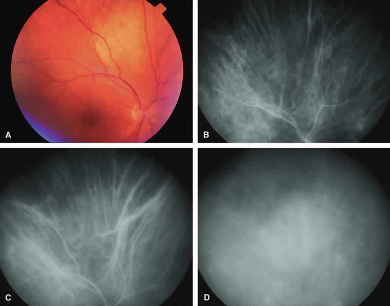
| Fig. 3. Amelanotic choroidal nevus. A. Ill-defined pale yellow choroidal nevus superior to optic disc. B-D. Indocyanine green (ICG) angiogram of lesion. B. Early-phase frame showing mild hypofluorescence of nevus and visibility of large-caliber choroidal arteries passing through lesion. C. Intermediate-phase frame showing features similar to those of the previous frame. D. Later phase frame showing ill-defined mild hyperfluorescence of lesion. |