|
|
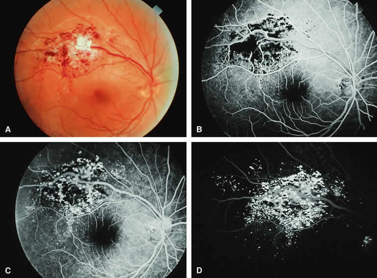
| Fig. 35. Retinal cavernous hemangioma. A. Cluster of dark red vascular saccules just superotemporal from center of macula along course of superotemporal branch retinal vein. White tissue on surface of lesion corresponds pathologically to gliosis. B-D. Fluorescein angiogram of lesion. B. Laminar venous phase angiogram showing abnormal retinal venous channels passing through largely hypofluorescent lesion. C. Full venous phase frame showing filling in of many vascular saccules comprising lesion. D. Late-phase frame showing hyperfluorescent cap of many component saccules of lesion, attributable to plasma-erythrocyte separation. |