|
|
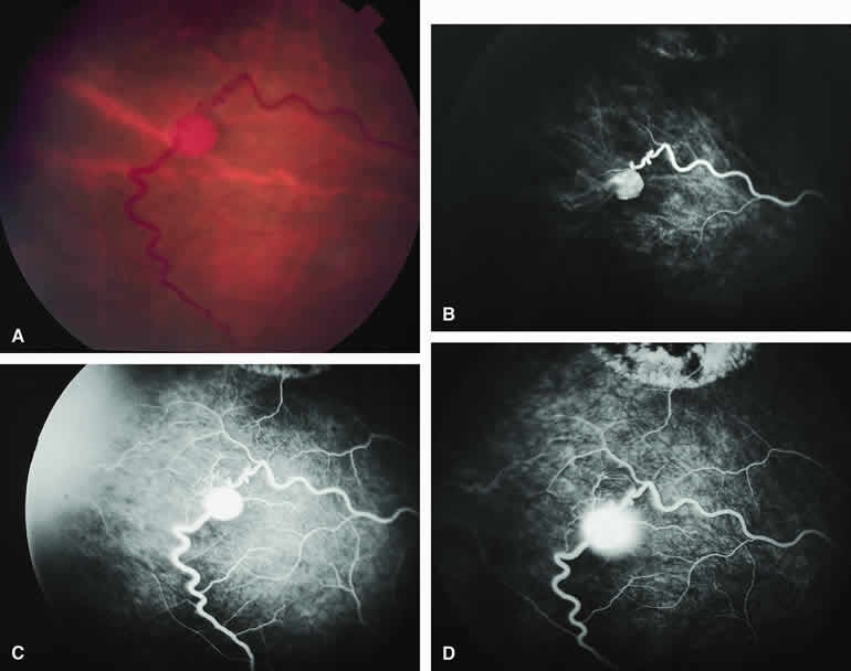
| Fig. 32. Small retinal capillary hemangioma. A. Spherical red vascular lesion in superotemporal periphery associated with prominent dilated, tortuous afferent and efferent retinal vascular channels. B-D. Fluorescein angiogram of lesion. B. Arterial phase frame showing filling of afferent arterial channel and lesion. C. Venous phase frame showing intense hyperfluorescence of vascular lesion, as well as complete filling of both afferent and efferent channels. D. Late-phase frame showing smudgy hyperfluorescence of lesion resulting from leakage of fluorescein into overlying vitreous and surrounding retina. Hypofluorescent and hyperfluorescent lesion at top of figure is site of a previously coagulated retinal capillary hemangioma. |