|
|
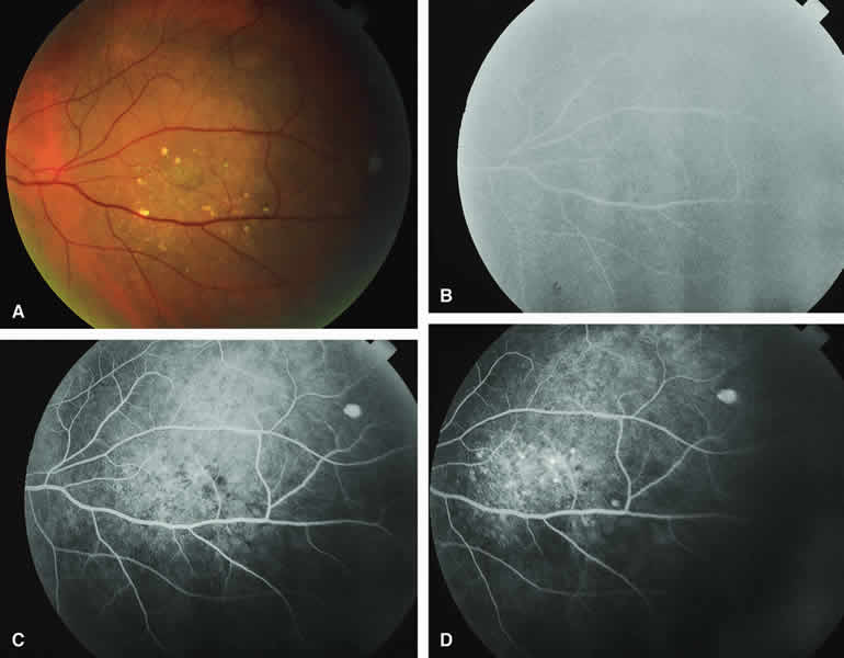
| Fig. 4. Melanotic choroidal nevus with drusen and retinal pigment epithelial (RPE) alterations. A. Gray-brown choroidal nevus with numerous white drusen and dark gray pigment clumps on its surface. B-D. Fluorescein angiogram of lesion. B. Venous phase frame showing retinal arteries and veins but no convincing sign of the underlying choroidal nevus. C. Later venous phase frame showing mild granular hyperfluorescence and hypofluorescence centrally corresponding with drusen and RPE pigment clumps overlying choroidal nevus. D. Late venous phase frame showing increased multifocal hyperfluorescence corresponding to drusen and persistent foci of hypofluorescence corresponding to RPE alternations. |