|
|
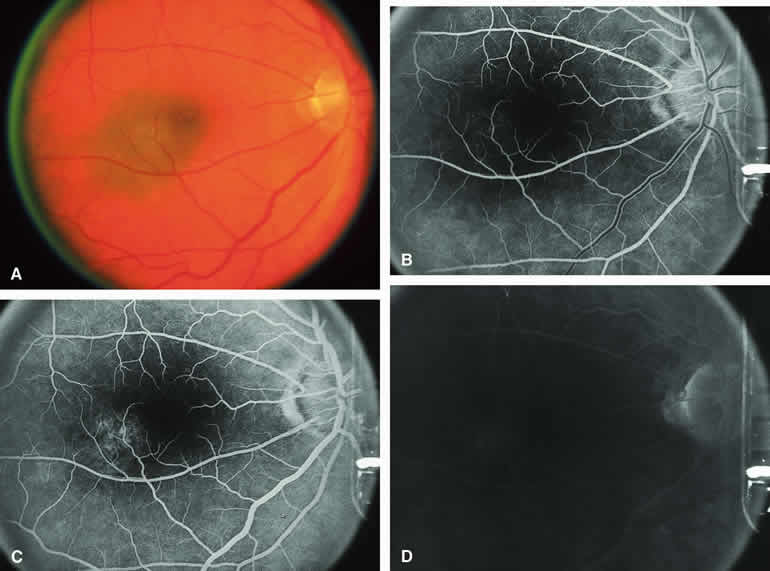
| Fig. 1. Typical melanotic choroidal nevus. A. Ill-defined gray-brown macular choroidal lesion with bland surface features. B-D. Fluorescein angiogram of lesion. B. Laminar venous phase frame showing intact retinal vasculature overlying nevus but virtually no hypofluorescence corresponding to choroidal lesion. C. Full venous phase frame showing granular hyperfluorescence corresponding to minor retinal pigment epithelial alterations and drusen on surface of tumor. D. Late-phase frame showing continued hypofluorescence of choroidal lesion with mild staining of surface drusen. |