|
|
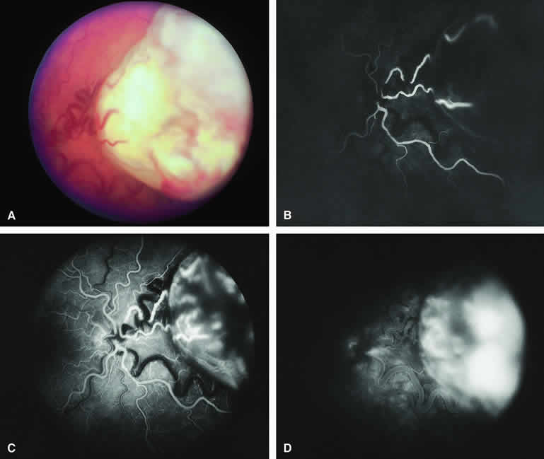
| Fig. 28. Larger intraretinal retinoblastoma with prominent retinal vasculature. A. Nodular white retinal tumor with associated dilated tortuous feeding retinal arteries and draining retinal veins. B-D. Fluorescein angiogram of lesions. B. Arterial phase frame showing rapid filling of retinal arteries feeding tumor. C. Early laminar venous phase frame showing fluorescence of large-caliber intralesional blood vessels. D. Late-phase frame showing diffuse hyperfluorescence of entire tumor and marked tortuosity of draining retinal veins. |