|
|
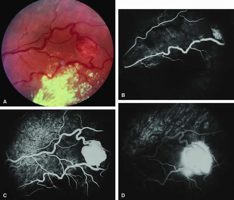
| Fig. 33. Larger retinal capillary hemangioma. A. Spherical pale red retinal lesion associated with dilated tortuous retinal arteries and veins and macular subretinal exudates. B-D. Fluorescein angiogram of lesion. B. Arterial phase frame showing dilation and tortuosity of feeding retinal artery and early hyperfluorescent filling of the vascular lesion. C. Venous phase frame showing intense hyperfluorescence of entire lesion plus fluorescein filling of the previously unfilled draining retinal vein. D. Late-phase frame showing smudgy leakage of fluorescein from the persistently hyperfluorescent retinal vascular lesion. |