|
|
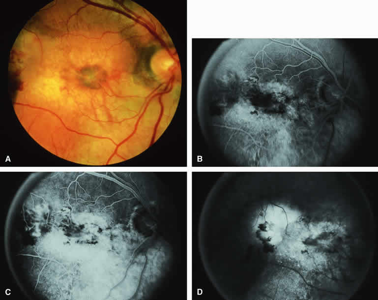
| Fig. 25. Choroidal osteoma with overlying choroidal neovascular membrane. A. Yellow-orange macular and juxtapapillary choroidal tumor with well-defined superior margin, prominent large-caliber intralesional blood vessels, a central macular black and red subretinal hemorrhagic figure, and bright red blood with turbid subretinal fluid at the left edge of the image. B-D. Fluorescein angiogram of lesion. B. Laminar venous phase frame showing generalized hyperfluorescence of choroidal lesion, variably intense hypofluorescence corresponding to the subretinal blood, and a partially well-defined choroidal neovascular membrane extending temporally from the central macula. C. Late laminar venous phase frame showing increased fluorescence of choroidal neovascular membrane and early smudgy leakage of fluorescein into overlying subretinal fluid. D. Late-phase frame showing persistent relative hyperfluorescence of choroidal mass, intense smudgy subretinal hyperfluorescence from the portion of the choroidal neovascular membrane at the temporal aspect of the macula, and sustained hypofluorescence corresponding to the subretinal blood. |