|
|
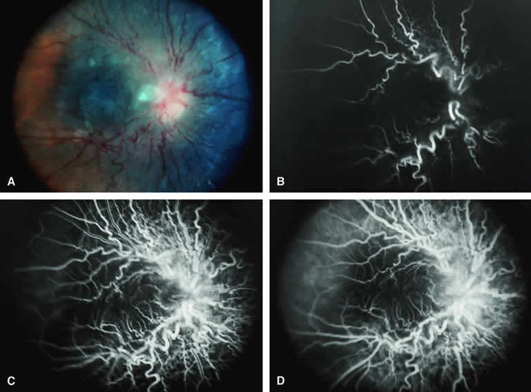
| Fig. 36. Combined hamartoma of retina. A. Ill-defined gray circumpapillary retinal lesion with whitish gliosis overlying portion of optic disc and prominent vascular tortuosity within lesion. B-D. Fluorescein angiogram of lesion. B. Arterial phase frame showing prominent tortuosity of large-caliber retinal blood vessels involved in mass. C. Laminar venous phase frame showing similar tortuosity of retinal veins and venules within lesion. D. Full venous phase frame showing ill-defined hypofluorescent background corresponding to darkly pigmented portion of mass. |