|
|
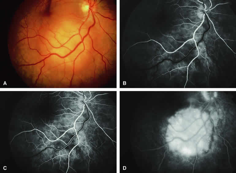
| Fig. 16. Typical amelanotic metastatic carcinoma to choroid. A. Yellow-white choroidal tumor located interotemporal from optic disc. Note associated turbid subretinal fluid (most evident in maculopapillary bundle region). B-D. Fluorescein angiogram of lesion. B. Laminar venous phase frame showing ill-defined mottled hypofluorescence and hyperfluorescence corresponding to clinically visible lesion. C. Later laminar venous phase frame still showing no distinct choroidal mass. D. Late-phase frame showing diffuse hyperfluorescence corresponding to lesion and leakage of fluorescein into subretinal space. |