|
|
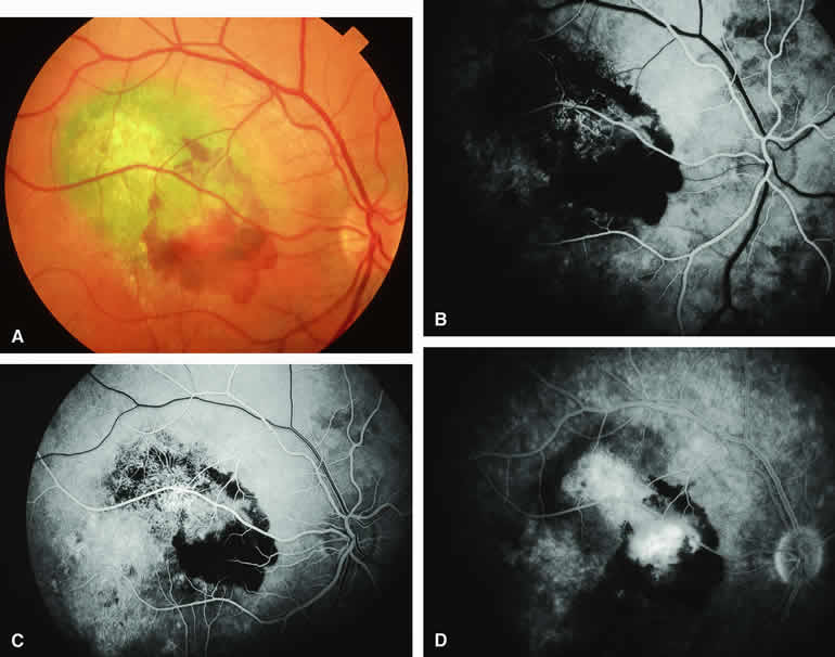
| Fig. 8. Melanotic choroidal nevus versus melanoma with overlying choroidal neovascular membrane. A. Same lesion shown in Figure 7 following spontaneous accumulation of subretinal blood and hard exudates associated with development of choroidal neovascular membrane overlying lesion. B-D. Fluorescein angiogram of lesion. B. Arterial phase frame showing abnormal vascular network deep to retina, partially obscured by blockage corresponding to subretinal blood. C. Laminar venous phase frame showing greater extent of abnormal vascular network overlying lesion and zone of window defect hyperfluorescence extending inferiorly through the macula. D. Late-phase frame showing smudgy hyperfluorescence from abnormal subretinal blood vessels, persistent blockage of choroidal fluorescence by the subretinal blood, and persistence of later hyperfluorescence due to retinal pigment epithelial (RPE) thinning extending inferiorly through the macula. |