|
|
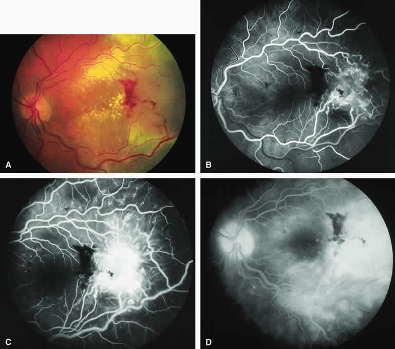
| Fig. 40. Amelanotic metastatic carcinoma to retina. A. Pale gray retinal infiltrate at temporal margin of photograph with associated intraretinal hemorrhages and hard exudates. The primary lesion was a cutaneous malignant melanoma. B-D. Fluorescein angiogram of lesion. B. Laminar venous phase frame showing abnormal retinal capillary network corresponding to tumor and fluorescence blockage corresponding to the retinal hemorrhages. C. Full venous phase frame showing intense smudgy hyperfluorescence of neoplastic retinal infiltrate. D. Late-phase frame showing intense expanded hyperfluorescence of the retinal lesion, diffuse hyperfluorescence of the surrounding retina and subretinal fluid, and persistent fluorescence blockage by the intraretinal blood. |