|
|
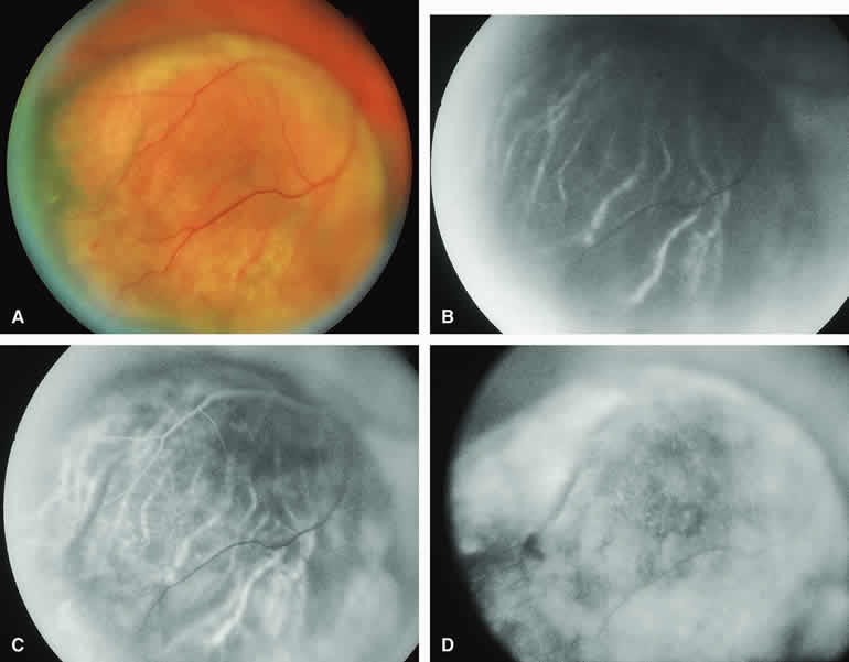
| Fig. 11. Amelanotic choroidal melanoma without invasive features. A. Amelanotic, golden-yellow choroidal tumor with prominent visibility of intralesional blood vessels. B-D. Fluorescein angiogram of lesion. B. Choroidal filling phase frame showing generalized hypofluorescence of lesion but with well-defined intralesional large-caliber blood vessels not connected to retinal vasculature. C. Retinal arterial phase frame showing even more prominent intralesional large blood vessels against persistently hypofluorescent tumor. D. Late-phase frame showing diffuse hyperfluorescence of surface of lesion resulting from exuberant leakage of fluorescein into subretinal space and lesion. |