|
|
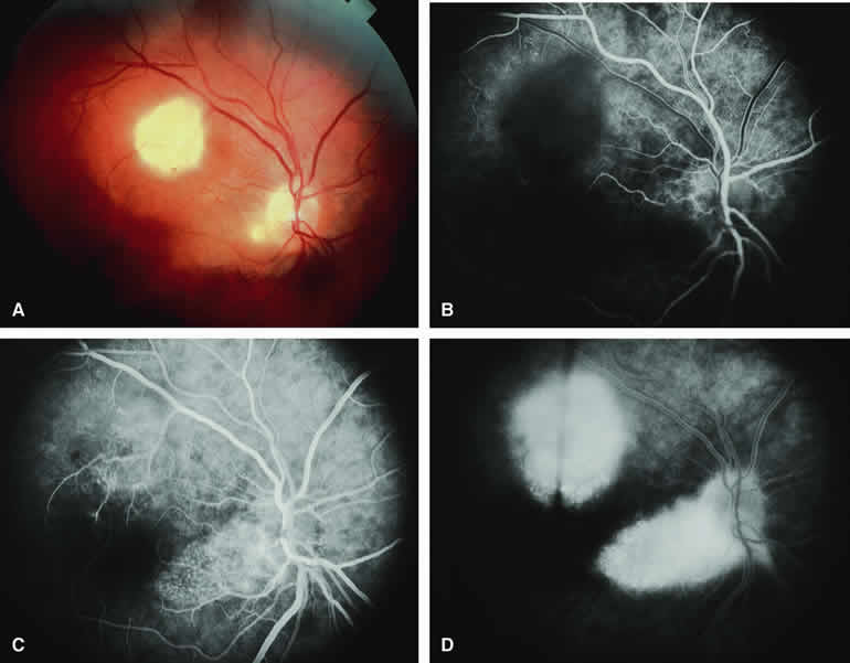
| Fig. 30. Multifocal retinal astrocytic hamartomas in tuberous sclerosis. A. Two white intraretinal lesions (larger lesion in superior macula, smaller lesion at inferotemporal margin of optic disc) in young adult patient with tuberous sclerosis. B-D. Fluorescein angiogram of lesions. B. Laminar venous phase fluorescein angiogram showing hypofluorescence of superior macular and inferotemporal maculopapillary bundle lesions. C. Venous phase frame showing smudgy hyperfluorescence corresponding to both retinal lesions. D. Late-phase frame showing diffuse staining of both lesions. |