|
|
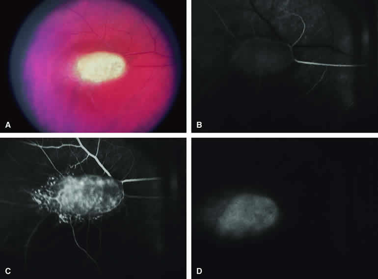
| Fig. 29. Retinal astrocytic hamartoma in child with tuberous sclerosis. A. Unifocal white inner retinal tumor without associated dilation or tortuosity of adjacent retinal arteries and veins. B-D. Fluorescein angiogram of lesion. B. Arterial phase frame showing faint pseudofluorescence of tumor and limited early filling of intralesional vasculature. C. Venous phase frame showing fluorescence of irregular capillary network within lesion. D. Late-phase frame showing staining of tumor. |