|
|
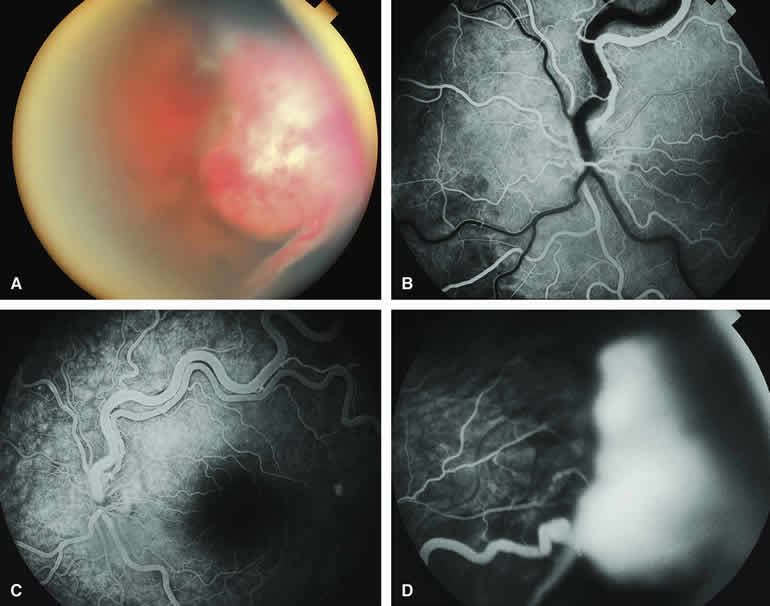
| Fig. 34. Relatively large peripheral retinal capillary hemangioma associated with subtotal tractional-exudative retinal detachment. A. Prominent red to white peripheral fundus lesion superotemporally associated with tractional-exudative retinal detachment. B-D. Fluorescein angiogram of lesion. B. Early laminar venous phase frame of posterior pole showing massive dilation and tortuosity of fluorescein-filled superotemporal branch retinal artery and unfilled superotemporal branch retinal vein. C. Venous phase frame of posterior pole showing fluorescein filling of both the superotemporal branch retinal artery and vein. D. Venous phase frame showing diffusely hyperfluorescent peripheral retinal lesion. |