|
|
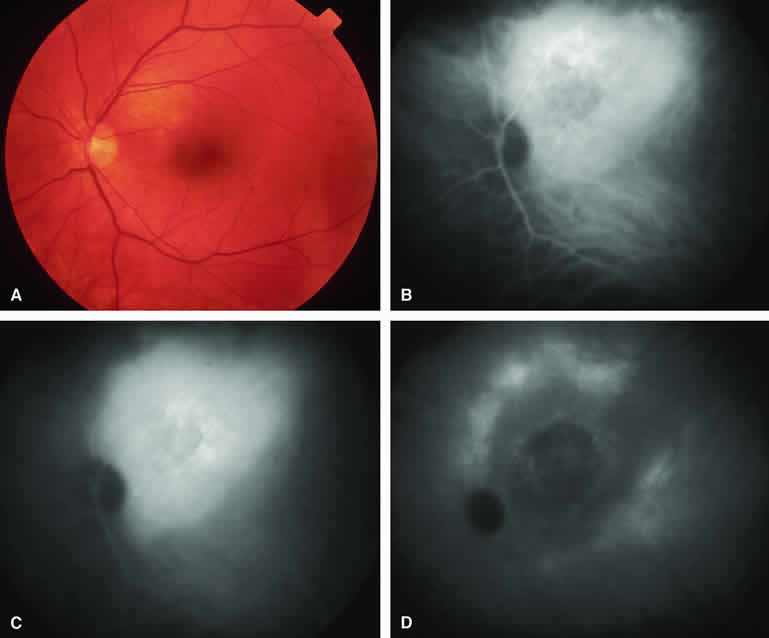
| Fig. 22. Circumscribed choroidal hemangioma. A. Ill-defined choroidal mass just superotemporal to optic disc with central pale zone corresponding to limited fibrous metaplasia of overlying retinal pigment epithelium. B-D. Indocyanine green (ICG) angiogram of lesion. B. Early-phase frame showing generalized hyperfluorescence of choroidal tumor. Note that basal extent of mass is much more apparent on angiogram than on color fundus photograph. C. Intermediate-phase frame showing persistent hyperfluorescence of choroidal mass. D. Very late phase (30 minutes postinjection) frame showing central washout of fluorescein with sustained marginal hyperfluorescence. |