|
|
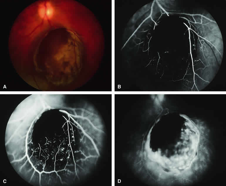
| Fig. 15. Choroidal melanoma with retinal invasion. A. Dark brown invasive tumor extends into retina and covers and obscures retinal blood vessels passing over tumor. B-D. Fluorescein angiogram of lesion. B. Laminar venous phase frame showing entire tumor to be hypofluorescent but with accentuated hypofluorescence corresponding to zone of retinal invasion. C. Full venous phase frame showing persistent intense hypofluorescence corresponding to zone of retinal invasion and alterations of adjacent retinal capillary bed indicative of retinal thinning and outer retinal invasion. D. Late-phase frame showing intense late hyperfluorescence of entire lesion with extension of fluorescein into subretinal space. Patch of retinal invasion remains hypofluorescent. |