|
|
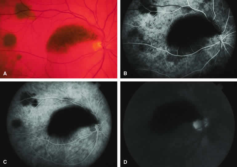
| Fig. 18. Melanotic metastatic cutaneous melanoma to choroid and retina. A. Melanotic metastatic lesions to choroid (three lesions deep to retinal vasculature at left side of photograph) and retina (arcuate lesion in papillomacular distribution). B-D. Fluorescein angiogram of lesions. B. Laminar venous phase frame showing complete hypofluorescence of retinal lesion and patchy partial hypofluorescence corresponding to three choroidal lesions. C. Full venous phase frame showing persistent well-defined hypofluorescence of retinal lesion, slightly increased choroidal hypofluorescence corresponding to the two larger choroidal lesions, and almost no abnormality corresponding to the smallest choroidal lesion. D. Late-phase frame showing persistent nonfluorescence of the retinal lesion but no abnormality corresponding to the three choroidal lesions. (Courtesy of Michael Bourne, MD) |