|
|
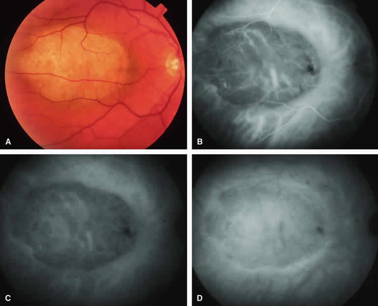
| Fig. 12. Amelanotic choroidal melanoma without invasive features. A. Pale yellow macular choroidal tumor with tiny clumps of disrupted retinal pigment epithelial (RPE) pigment on its surface. B-D. Indocyanine green (ICG) angiogram of lesion. B. Early-phase frame showing prominent fluorescent large-caliber intralesional blood vessels against the background of a generally hypofluorescent choroidal mass. C. Later phase frame showing persistent hypofluorescence of the mass except for the prominent intralesional blood vessels. D. Late-phase frame showing increase in fluorescence of mass compared with surrounding normal choroid and marginal hyperfluorescence resulting from ICG accumulation in the overlying subretinal fluid. |